iBet uBet web content aggregator. Adding the entire web to your favor.


Link to original content: https://pubmed.ncbi.nlm.nih.gov/20699274
TAL nucleases (TALNs): hybrid proteins composed of TAL effectors and FokI DNA-cleavage domain - PubMed Skip to main page content
U.S. flag

An official website of the United States government

Dot gov

The .gov means it’s official.
Federal government websites often end in .gov or .mil. Before sharing sensitive information, make sure you’re on a federal government site.

Https

The site is secure.
The https:// ensures that you are connecting to the official website and that any information you provide is encrypted and transmitted securely.

Access keys NCBI Homepage MyNCBI Homepage Main Content Main Navigation
. 2011 Jan;39(1):359-72.
doi: 10.1093/nar/gkq704. Epub 2010 Aug 10.

TAL nucleases (TALNs): hybrid proteins composed of TAL effectors and FokI DNA-cleavage domain

Affiliations

TAL nucleases (TALNs): hybrid proteins composed of TAL effectors and FokI DNA-cleavage domain

Ting Li et al. Nucleic Acids Res. 2011 Jan.

Abstract

DNA double-strand breaks enhance homologous recombination in cells and have been exploited for targeted genome editing through use of engineered endonucleases. Here we report the creation and initial characterization of a group of rare-cutting, site-specific DNA nucleases produced by fusion of the restriction enzyme FokI endonuclease domain (FN) with the high-specificity DNA-binding domains of AvrXa7 and PthXo1. AvrXa7 and PthXo1 are members of the transcription activator-like (TAL) effector family whose central repeat units dictate target DNA recognition and can be modularly constructed to create novel DNA specificity. The hybrid FN-AvrXa7, AvrXa7-FN and PthXo1-FN proteins retain both recognition specificity for their target DNA (a 26 bp sequence for AvrXa7 and 24 bp for PthXo1) and the double-stranded DNA cleaving activity of FokI and, thus, are called TAL nucleases (TALNs). With all three TALNs, DNA is cleaved adjacent to the TAL-binding site under optimal conditions in vitro. When expressed in yeast, the TALNs promote DNA homologous recombination of a LacZ gene containing paired AvrXa7 or asymmetric AvrXa7/PthXo1 target sequences. Our results demonstrate the feasibility of creating a tool box of novel TALNs with potential for targeted genome modification in organisms lacking facile mechanisms for targeted gene knockout and homologous recombination.


Figures

Figure 1.
Figure 1.
Schematics of TAL effectors AvrXa7 and PthXo1 as well as their fusions with FN. (A) A typical TAL effector contains a central region of 34 aa direct repeats (open boxes) and three nuclear localization motifs (NLS, black bars) as well as a transcription activation domain (AD, red solid box) at the C-terminus. The representative 34 aa repeat is shown below with the variable amino acid residues at the positions 12 and 13 in red. (B) and (C) AvrXa7 and PthXo1 contain central 26 and 24 tandem repeats (shown as boxes) of 34 aa residues, respectively. The repeats are highly conserved, except for residues at positions 12 and 13 (asterisk, missing amino acid residue at position 13). The sequence recognition of AvrXa7 to Os11N3 (and PthXo1 to Os8N3) is dictated by the order of the 34 aa repeat units that each contain at positions 12 and 13, a pair of amino acids that exactly recognize one nucleotide at one position in the DNA sequence of the EBE of the Os11N3 gene. (D–F) Schematics of TAL effector fusion proteins with FN domain at the N-terminus of AvrXa7 (D) or at the C-termini of AvrXa7 (E) and PthXo1 (F). (G) Transient activation by AvrXa7 and FN-AvrXa7 of the Os11N3 promoter (containing the AvrXa7 EBE) driving a GFP reporter gene (GFP-EBE). The plasmid containing the GFP reporter gene was co-injected along with a plasmid containing a gene encoding the AvrXa7 or AvrXa7-FN driven by the CaMV 35S promoter [or with a plasmid containing the 35S promoter but no AvrXa7 coding sequence (Control)]. The dotted line indicates the edge of the inoculation site.
Figure 2.
Figure 2.
Binding specificity of AvrXa7-FN and FN-AvrXa7 fusion proteins to their target DNA. (A) Sense-strand sequences of the wild type (Os11N3) and mutant (Os11N3 M) oligonucleotide duplexes used in the EMS assays. (B) EMS assays demonstrating specificities of AvrXa7-FN and FN-AvrXa7 binding to the AvrXa7 EBE sequence. The set of gel images to the left depict results with AvrXa7-FN, while set of gel images to the right depict results with FN-AvrXa7. The left panels of each set show binding of AvrXa7-FN and FN-AvrXa7 to the authentic, 32P-labeled, Os11N3 DNA target element but not to the mutated target. Competition assays (middle and right panels of each set) showing that the binding of 32P-labeled Os11N3 EBE target by AvrXa7-FN and FN-AvrXa7 is effectively competed by excess amounts of non-radioactive Os11N3 oligonucleotides (middle panels of each set), but not by the non-radioactive mutated version of Os11N3 DNA (right panels). Positions of the bound and free probes are indicated at the left of the autoradiograph.
Figure 3.
Figure 3.
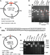
Target DNA digestion by the FN-AvrXa7, AvrXa7-FN and PthXo1-FN TALNs. (A) Circular plasmid containing a 400 bp Os11N3 promoter region (thick bar in red) with one AvrXa7 EBE site (position 0). EcoNI cuts once at position 842 and MluI cuts twice at positions 1405 and 2246 relative to EBE of the plasmid. The expected sizes of fragments after cleavages are shown below the plasmid map. (B) Gel image of plasmid DNA treated with different enzymes individually or in combinations. M, 1 kb markers with size labeled on the left side. Different treatments are indicated above lanes 1–9 and in combination with TALNs labeled below lanes 4–9. Arrows in red indicate DNA fragment sizes expected if the NF-AvrXa7 or AvrXa7-FN nuclease cleavage takes place correctly at the AvrXa7 EBE target site in pTOP/Os11N3. (C) Structure of adjacent PthXo1 and AvrXa7 EBE sites (underlined) in a TD (to allow FN to FN proximity) and separated by 19 spacer nucleotides (lower case letters) which contains a unique EcoRI site (underlined). (D) Map of plasmid pEBE-DT with predicted cleavage sites for EcoRI (position 0), PthXo1-FN and/or AvrXa7-FN EBEs and BglI (positions 1479 and 2745). (E) Gel image of intact pEBE-DT (lane 1), EcoRI–linearized plasmid (2), BglI-cleaved plasmid (3), AvrXa7-FN digested plasmid DNA (4), AvrXa7-FN digested plasmid followed by BglI digestion (5), PthXo1-FN digested plasmid (6), PthXo1-FN digested plasmid followed by BglI digestion (7) and double digestion with PthXo1-FN and AvrXa7-FN (at one-fifth the concentrations of each protein used individually in reactions for lanes 3 to 6) followed by BglI digestion (8). The label ‘M’ indicates the DNA marker lane with marker sizes provided in kilo-base (kb). Arrows to the right indicate sizes of DNA fragments expected from complete DNA digestion with BglI and partial digestion with PthXo1-FN (lane 5), AvrXa7-FN (lane 7) or PthXo1-FN combined with AvrXa7-FN (lane 8).
Figure 4.
Figure 4.
DNA sequencing revealing the major cleavage sites created by the AvrXa7-FN (A) and FN-AvrXa7 (B). (A) DNA sequencing chromatogram above the original Os11N3 dsDNA sequence (sense strand colored for ease of viewing) is based on the sense strand of the 0.8 kb fragment purified from lane 8 in Figure 3B. The M13 R primer was used for sequencing the sense strand. The chromatogram, which represents the sense strand sequence around the cleavage site, is in reverse-complement orientation for ease of viewing. The chromatogram below the dsDNA sequence is derived from the antisense strand of the 2.1 kb DNA fragment (also from lane 8 in Figure 3B) to the left of the predicted AvrXa7-FN cleavage site. The AvrXa7-FN binding site is boxed in shaded gray. The vertical arrows denote the obvious cleavage sites in both strands. The thick underline indicates the 15 bp region downstream of the EBE site in which the vast majority of AvrXa7-FN cleavage occurred. (B) DNA sequencing chromatograms of two DNA fragments derived from pTOP/11N3 treated with FN-AvrXa7 and purified from lane 5 in Figure 3B. Labeling is similar to that used for (A).
Figure 5.
Figure 5.
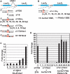
Yeast SSA assay for detection of FN-AvrXa7, AvrXa7-FN and PthXo1-FN TALN-induced HR. (A) Schematics of the reporter constructs (not drawn to scale) with dual BCR-ABL target site, AvrXa7 EBE site in different configurations drawn below. Two non-functional LacZ gene fragments (LacZn and LacZc, blue solid bars) were separated by the yeast URA3 gene (gray line) and a MCS (black line). The 125 bp duplicated LacZ coding sequences are represented as hatched blue boxes. The target sites in the reporter constructs are designated as zf-TD6 (ZFN dual sites), x7-S (single EBE site), x7-DHn1 (dual AvrXa7 EBEs in a HD separated by the red-lined spacers), x7-TDn2 (dual tail-to-tail AvrXa7 EBEs) followed by the number of spacer nucleotides. The constructs x7-TD19m1 and x7-TD19m2 contain a 19 bp spacer and carry a mutation in their EBE site caused by a 4 bp substitution (depicted as a red cross). (B) Schematics similar to (A) with a 125 bp fragment (gray line) instead of the URA3 gene between the MCS and LacZn. (C) Activity of FN-AvrXa7 on reporters with different lengths of spacer between the AvrXa7 EBE sites (respective spacer lengths are given below each column). Fold change represents the β-galactosidase activity of yeast cells co-expressing the respective reporter and FN-AvrXa7 compared to cells containing the same reporter but lacking the FN-AvrXa7 expression vector. (D) Stimulation by the ZFN, AvrXa7-FN and AvrXa7-FN/pthXo1-FN constructs of their respective reporter genes containing various spacers (lengths in base pairs given below each column). Fold change represents the β-galactosidase activity of yeast cells co-expressing each reporter and its respective effector nuclease(s) (indicated below the columns) compared to those containing the same reporter but lacking the presence of genes encoding the respective nuclease(s). Error bars represent the standard deviation of three samples.

Similar articles

Cited by

References

    1. Le Provost F, Lillico S, Passet B, Young R, Whitelaw B, Vilotte JL. Zinc finger nuclease technology heralds a new era in mammalian transgenesis. Trends Biotechnol. 2010;28:134–141. - PubMed
    1. Jasin M. Genetic manipulation of genomes with rare-cutting endonucleases. Trends Genet. 1996;12:224–228. - PubMed
    1. Vasquez KM, Marburger K, Intody Z, Wilson JH. Manipulating the mammalian genome by homologous recombination. Proc. Natl Acad. Sci. USA. 2001;98:8403–8410. - PMC - PubMed
    1. Bibikova M, Beumer K, Trautman JK, Carroll D. Enhancing gene targeting with designed zinc finger nucleases. Science. 2003;300:764. - PubMed
    1. Porteus MH, Carroll D. Gene targeting using zinc finger nucleases. Nat. Biotechnol. 2005;23:967–973. - PubMed

Publication types

MeSH terms

Substances