Stage 3 immature human natural killer cells found in secondary lymphoid tissue constitutively and selectively express the TH 17 cytokine interleukin-22
- PMID: 19244159
- PMCID: PMC2673127
- DOI: 10.1182/blood-2008-12-192443
Stage 3 immature human natural killer cells found in secondary lymphoid tissue constitutively and selectively express the TH 17 cytokine interleukin-22
Abstract
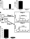
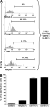
Considerable functional heterogeneity within human natural killer (NK) cells has been revealed through the characterization of distinct NK-cell subsets. Accordingly, a small subset of CD56(+)NKp44(+)NK cells, termed NK-22 cells, was recently described within secondary lymphoid tissue (SLT) as IL-22(-) when resting, with a minor fraction of this population becoming IL-22(+) when activated. Here we discover that the vast majority of stage 3 immature NK (iNK) cells in SLT constitutively and selectively express IL-22, a T(H)17 cytokine important for mucosal immunity, whereas earlier and later stages of NK developmental intermediates do not express IL-22. These iNK cells have a surface phenotype of CD34(-)CD117(+)CD161(+)CD94(-), largely lack expression of NKp44 and CD56, and do not produce IFN-gamma or possess cytolytic activity. In summary, stage 3 iNK cells are highly enriched for IL-22 and IL-26 messenger RNA, and IL-22 protein production, but do not express IL-17A or IL-17F.
Figures


Similar articles
-
Interleukin-1beta selectively expands and sustains interleukin-22+ immature human natural killer cells in secondary lymphoid tissue.Immunity. 2010 Jun 25;32(6):803-14. doi: 10.1016/j.immuni.2010.06.007. Immunity. 2010. PMID: 20620944 Free PMC article.
-
The abundant NK cells in human secondary lymphoid tissues require activation to express killer cell Ig-like receptors and become cytolytic.J Immunol. 2004 Feb 1;172(3):1455-62. doi: 10.4049/jimmunol.172.3.1455. J Immunol. 2004. PMID: 14734722
-
Development of IL-22-producing NK lineage cells from umbilical cord blood hematopoietic stem cells in the absence of secondary lymphoid tissue.Blood. 2011 Apr 14;117(15):4052-5. doi: 10.1182/blood-2010-09-303081. Epub 2011 Feb 10. Blood. 2011. PMID: 21310921 Free PMC article.
-
Human natural killer cell development in secondary lymphoid tissues.Semin Immunol. 2014 Apr;26(2):132-7. doi: 10.1016/j.smim.2014.02.008. Epub 2014 Mar 21. Semin Immunol. 2014. PMID: 24661538 Free PMC article. Review.
-
Interleukin-22-producing natural killer cells and lymphoid tissue inducer-like cells in mucosal immunity.Immunity. 2009 Jul 17;31(1):15-23. doi: 10.1016/j.immuni.2009.06.008. Immunity. 2009. PMID: 19604490 Review.
Cited by
-
Cellular Origins and Pathogenesis of Gastrointestinal NK- and T-Cell Lymphoproliferative Disorders.Cancers (Basel). 2022 May 18;14(10):2483. doi: 10.3390/cancers14102483. Cancers (Basel). 2022. PMID: 35626087 Free PMC article. Review.
-
Innate lymphoid cells: NK and cytotoxic ILC3 subsets infiltrate metastatic breast cancer lymph nodes.Oncoimmunology. 2022 Mar 30;11(1):2057396. doi: 10.1080/2162402X.2022.2057396. eCollection 2022. Oncoimmunology. 2022. PMID: 35371620 Free PMC article.
-
Modulation of Intestinal ILC3 for the Treatment of Type 1 Diabetes.Front Immunol. 2021 Jun 3;12:653560. doi: 10.3389/fimmu.2021.653560. eCollection 2021. Front Immunol. 2021. PMID: 34149694 Free PMC article. Review.
-
The Influence of Innate Lymphoid Cells and Unconventional T Cells in Chronic Inflammatory Lung Disease.Front Immunol. 2019 Jul 11;10:1597. doi: 10.3389/fimmu.2019.01597. eCollection 2019. Front Immunol. 2019. PMID: 31354734 Free PMC article. Review.
-
IL-26 contributes to host defense against intracellular bacteria.J Clin Invest. 2019 Apr 2;129(5):1926-1939. doi: 10.1172/JCI99550. eCollection 2019 Apr 2. J Clin Invest. 2019. PMID: 30939123 Free PMC article.
References
-
- Freud AG, Becknell B, Roychowdhury S, et al. A human CD34(+) subset resides in lymph nodes and differentiates into CD56bright natural killer cells. Immunity. 2005;22:295–304. - PubMed
-
- Wolk K, Kunz S, Witte E, Friedrich M, Asadullah K, Sabat R. IL-22 increases the innate immunity of tissues. Immunity. 2004;21:241–254. - PubMed
MeSH terms
Substances
LinkOut - more resources
Full Text Sources
Other Literature Sources
Research Materials


