iBet uBet web content aggregator. Adding the entire web to your favor.


Link to original content: https://pubmed.ncbi.nlm.nih.gov/18816377/
The chemokine CXCL1/growth related oncogene increases sodium currents and neuronal excitability in small diameter sensory neurons - PubMed Skip to main page content
U.S. flag

An official website of the United States government

Dot gov

The .gov means it’s official.
Federal government websites often end in .gov or .mil. Before sharing sensitive information, make sure you’re on a federal government site.

Https

The site is secure.
The https:// ensures that you are connecting to the official website and that any information you provide is encrypted and transmitted securely.

Access keys NCBI Homepage MyNCBI Homepage Main Content Main Navigation
. 2008 Sep 24:4:38.
doi: 10.1186/1744-8069-4-38.

The chemokine CXCL1/growth related oncogene increases sodium currents and neuronal excitability in small diameter sensory neurons

Affiliations

The chemokine CXCL1/growth related oncogene increases sodium currents and neuronal excitability in small diameter sensory neurons

Jun-Gang Wang et al. Mol Pain. .

Abstract

Background: Altered Na+ channel expression, enhanced excitability, and spontaneous activity occur in nerve-injury and inflammatory models of pathological pain, through poorly understood mechanisms. The cytokine GRO/KC (growth related oncogene; CXCL1) shows strong, rapid upregulation in dorsal root ganglion in both nerve injury and inflammatory models. Neurons and glia express its receptor (CXCR2). CXCL1 has well-known effects on immune cells, but little is known about its direct effects on neurons.

Results: We report that GRO/KC incubation (1.5 nM, overnight) caused marked upregulation of Na+ currents in acutely isolated small diameter rat (adult) sensory neurons in vitro. In both IB4-positive and IB4-negative sensory neurons, TTX-resistant and TTX-sensitive currents increased 2- to 4 fold, without altered voltage dependence or kinetic changes. These effects required long exposures, and were completely blocked by co-incubation with protein synthesis inhibitor cycloheximide. Amplification of cDNA from the neuronal cultures showed that 3 Na channel isoforms were predominant both before and after GRO/KC treatment (Nav 1.1, 1.7, and 1.8). TTX-sensitive isoforms 1.1 and 1.7 significantly increased 2 - 3 fold after GRO/KC incubation, while 1.8 showed a trend towards increased expression. Current clamp experiments showed that GRO/KC caused a marked increase in excitability, including resting potential depolarization, decreased rheobase, and lower action potential threshold. Neurons acquired a striking ability to fire repetitively; IB4-positive cells also showed marked broadening of action potentials. Immunohistochemical labelling confirmed that the CXCR2 receptor was present in most neurons both in dissociated cells and in DRG sections, as previously shown for neurons in the CNS.

Conclusion: Many studies on the role of chemokines in pain conditions have focused on their rapid and indirect effects on neurons, via release of inflammatory mediators from immune and glial cells. Our study suggests that GRO/KC may also have important pro-nociceptive effects via its direct actions on sensory neurons, and may induce long-term changes that involve protein synthesis.


Figures
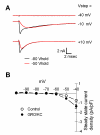
Figure 1
Figure 1
GRO/KC incubated cells have Na+ currents that are qualitatively similar to those seen in control cells. A. Examples of current traces elicited from holding potential of -80 mV (darker, smaller traces) or -50 mV. As in control cells, the currents elicited from -80 mV have a faster component, which is TTX-sensitive (see text). B. Persistent Na+ currents activating at -60 to -50 mV were not observed. Average leak-subtracted steady state current is plotted against the step potential. Holding potential was -80 mV.
Figure 2
Figure 2
Overnight incubation with GRO/KC (1.5 nM) increases Na+ current density. A. TTX-R current density significantly increased in IB4-negative cells after GRO/KC incubation. P < 0.0001 for overall effect. Individual voltages at which the difference between control and GRO/KC treated cells was significant are indicated by the * symbol (two-way RM ANOVA with Holm-Sidak method for Pairwise Multiple Comparisons). B. A similar but smaller enhancement of TTX-R current was observed in IB4-positive cells (overall p value = 0.0016). TTX-S current density measured at -30 to -10 mV as described in methods, also significantly increased after GRO/KC incubation in both IB4-negative (C; and IB4-positive cells (D). Data are from 9 cultures; N = 53 control cells and 54 GRO/KC treated cells.
Figure 3
Figure 3
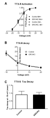
Overnight incubation with GRO/KC does not affect decay time constants or activation of Na+ currents. A. Activation data for the TTX-R current was fit by a standard Boltzmann equation. No differences were found in the fit parameters between the 4 experimental groups (p = 0.22). The plotted line represents the best fit to all the data, and has a V1/2 value of -7.3 mV and a slope factor of 5.7. B. Time constants for the decay of TTX-R current were obtained by fitting single exponentials to the falling phase of currents evoked from a holding potential of -50 mV. No significant difference between the two groups was observed (two-way RM ANOVA). C. Time constants for the TTX-S current at were obtained by fitting the decaying phase of the current with the sum of two exponentials. The slower of these corresponded to the time constant observed in the TTX-R current, and the faster time constant was used as the value of the TTX-S decay time constant. No significant difference between the two groups was observed (Mann-Whitney test, p = 0.18). Data from IB4-positive and IB4-negative cells were combined as no difference between these groups in decay time constants was observed.
Figure 4
Figure 4
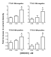
Lower doses of GRO/KC do not increase Na+ current density. Current densities were measured as in Figure 1, after overnight exposure to the indicated concentration of GRO/KC. For each concentration tested, data have been normalized to the control values obtained in cells from the same cultures with no GRO/KC treatment. *, significantly different from control. TTX-R current at 0 mV was used for this analysis. N = 27 cells for 0.06 nM dose (plus 17 control cells from the same cultures); 10 cells for 0.28 nM dose (plus 14 control cells), and 54 cells for 1.5 nM dose (plus 53 control cells).
Figure 5
Figure 5
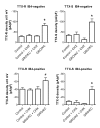
Effect of the protein synthesis inhibitor cycloheximide (CHX) on enhancement of Na+ currents by GRO/KC incubation. Cycloheximide (3.5 μM) or vehicle (DMSO) was added to cell cultures just before addition of 1.5 nM GRO/KC or vehicle, and TTX-S and TTX-R Na currents were measured 16 to 30 hours later. *, significantly different from all other groups; #, significantly different from control (one-way ANOVA followed by Tukey's multiple comparison test). Data are from 1 set of experiments (3 cultures) comparing 22 control and 29 CHX treated cells, and a second set of experiments (2 cultures) comparing 14 GRO/KC treated cells with 22 GRO/KC + CHX treated cells. Additional data, from Figure 1, are included in the control and GRO/KC data groups. Analysis omitting this additional data gave similar results, except that the TTX-R current in IB4-positive cells showed no significant differences between groups.
Figure 6
Figure 6
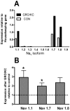
Expression of Na channels Nav1.1 through Nav 1.9 in cultured DRG cells before and after GRO/KC incubation. cDNA template was reverse-transcribed from RNA that had been isolated from control cells and cells from the sister cultures treated overnight with GRO/KC (1.5 nM). Expression of each gene was normalized to that of the housekeeping gene HPRT in the same batch of cDNA, determined during the same PCR amplification. A, profile of Na channel expression relative to HPRT. B, fold-change in expression of the 3 highly expressed Na channels after GRO/KC treatment. *, significantly different from control (= 1.0, dotted line), ratio t-test. Values shown are averages from 4 separate cultures.
Figure 7
Figure 7
Small diameter neurons acquire the ability to fire repetitively after GRO/KC incubation. A, Examples of the voltage response to a 90 pA 1 second current injection in a control IB4-negative neuron (top) and in an IB4-negative neuron incubated overnight in GRO/KC (1.5 nM) (bottom). B. Examples of the voltage response to a 50 pA current injection in control (top) and GRO/KC incubated (bottom) IB4-positive neurons. Same scale as A. C, D: Average number of action potentials during a 1 second current injection as a function of current amplitude in IB4-negative (C) and IB4-positive (D) cells. *, significant difference between GRO/KC and control cells at the indicated current value (two-way RM ANOVA with Holm-Sidak post test). N = 8 to 14 cells per group; data combined from 3 different cultures.
Figure 8
Figure 8
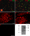
Immunohistochemical detection and immunofluorescence double labelling of CXCR2 in acutely dissociated DRG neurons and in DRG sections from rat. A: Double staining of the dissociated DRG cells with anti-CXCR2 and anti-NeuN showing that high CXCR2-expressing cells are neuronal cells, and that the more modest staining levels observed occur in almost all DRG neurons. B: Neurons with intense CXCR2 staining are all IB4-negative. C: CXCR2 staining in DRG sections showing expression on plasma membrane of most neurons, of small, medium and large diameters, with high levels expression in some smaller neurons. CXCR2 is also expressed in the nuclear membrane in some cells (arrows), as can be seen more clearly in the higher magnification view in D. E: Negative control. DRG sections incubated with primary antibody preabsorbed with 30 fold excess of antigen did not show any immunoreactivity. Scale bar = 50 μm. F. Western blot of protein isolated from DRG neurons, using the same antibody (1:300) as a probe. Left panel: standard. Right panel: CXCR2.

Similar articles

Cited by

References

    1. Cummins TR, Sheets PL, Waxman SG. The roles of sodium channels in nociception: Implications for mechanisms of pain. Pain. 2007;131:243–257. doi: 10.1016/j.pain.2007.07.026. - DOI - PMC - PubMed
    1. Sommer C, Kress M. Recent findings on how proinflammatory cytokines cause pain: peripheral mechanisms in inflammatory and neuropathic hyperalgesia. Neurosci Lett. 2004;361:184–187. doi: 10.1016/j.neulet.2003.12.007. - DOI - PubMed
    1. Moalem G, Grafe P, Tracey DJ. Chemical mediators enhance the excitability of unmyelinated sensory axons in normal and injured peripheral nerve of the rat. Neuroscience. 2005;134:1399–1411. doi: 10.1016/j.neuroscience.2005.05.046. - DOI - PubMed
    1. Watkins LR, Milligan ED, Maier SF. Glial proinflammatory cytokines mediate exaggerated pain states: implications for clinical pain. Adv Exp Med Biol. 2003;521:1–21. - PubMed
    1. DeLeo JA, Tanga FY, Tawfik VL. Neuroimmune activation and neuroinflammation in chronic pain and opioid tolerance/hyperalgesia. Neuroscientist. 2004;10:40–52. doi: 10.1177/1073858403259950. - DOI - PubMed

Publication types

LinkOut - more resources