iBet uBet web content aggregator. Adding the entire web to your favor.


Link to original content: https://pubmed.ncbi.nlm.nih.gov/18323453/
De novo computational design of retro-aldol enzymes - PubMed Skip to main page content
U.S. flag

An official website of the United States government

Dot gov

The .gov means it’s official.
Federal government websites often end in .gov or .mil. Before sharing sensitive information, make sure you’re on a federal government site.

Https

The site is secure.
The https:// ensures that you are connecting to the official website and that any information you provide is encrypted and transmitted securely.

Access keys NCBI Homepage MyNCBI Homepage Main Content Main Navigation
. 2008 Mar 7;319(5868):1387-91.
doi: 10.1126/science.1152692.

De novo computational design of retro-aldol enzymes

Affiliations

De novo computational design of retro-aldol enzymes

Lin Jiang et al. Science. .

Abstract

The creation of enzymes capable of catalyzing any desired chemical reaction is a grand challenge for computational protein design. Using new algorithms that rely on hashing techniques to construct active sites for multistep reactions, we designed retro-aldolases that use four different catalytic motifs to catalyze the breaking of a carbon-carbon bond in a nonnatural substrate. Of the 72 designs that were experimentally characterized, 32, spanning a range of protein folds, had detectable retro-aldolase activity. Designs that used an explicit water molecule to mediate proton shuffling were significantly more successful, with rate accelerations of up to four orders of magnitude and multiple turnovers, than those involving charged side-chain networks. The atomic accuracy of the design process was confirmed by the x-ray crystal structure of active designs embedded in two protein scaffolds, both of which were nearly superimposable on the design model.


Figures
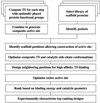
Fig. 1
Fig. 1
Computational enzyme design protocol for a multistep reaction. The first step is to generate ensembles of models of each of the key intermediates and transition states in the reaction pathway in the context of a specific catalytic motif composed of protein functional groups. These models are then superimposed, based on the protein functional group positions, to create an initial composite active-site description. Large ensembles of distinct 3D realization of these composite active sites are then generated by simultaneously varying the degrees of freedom of the composite TS, the orientation of the catalytic side chains relative to the composite TS, and the internal conformation of the catalytic side chains. For each composite active-site description, candidate catalytic sites are generated in an input scaffold set by RosettaMatch (15). Briefly, each rotamer of each catalytic side chain is placed at each position in each scaffold, and the ensuing position of the composite TS is recorded in the hash. After the filling out of the hash table, which is linear in the numbers of scaffold positions and catalytic rotamers, the hash is searched for TS positions that are compatible with all catalytic constraints; such positions are termed “matches.” For each match, the rigid body orientation of the composite TS and the internal coordinates of the catalytic side chains are optimized to reduce steric clashes while maintaining the catalytic geometry within specified tolerances. The remaining positions (not included in the minimal catalytic site description) surrounding the docked composite TS model are redesigned to optimize TS binding affinity by means of the standard Rosetta design methodology (20, 21). The rigid body orientation of the composite TS, the side-chain torsion angles, and (in some cases) the backbone torsion angles in the active site are refined via quasi-Newton optimization (22). The resulting designs are ranked based on the total binding energy to the composite TS and the satisfaction of the specified catalytic geometry, and then the top-ranked designs are experimentally characterized. The SOM contains detailed descriptions of each step in the protocol.
Fig. 2
Fig. 2
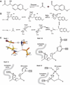
Retro-aldol reaction and active-site motifs. (A) The retro-aldol reaction. (B) General description of the aldol reaction pathway with a nucleophilic lysine and general acid-base chemistry. Several of the proton transfer steps are left out for brevity. (C) Active-site motifs with quantum mechanically optimized structures (23). (Top left) Motif I. Two lysines are positioned nearby one another to lower the pKa of the nucleophilic lysine, and a Lys-Asp dyad acts as the base to deprotonate the hydroxyl group. (Bottom left) Motif II. The catalytic lysine is buried in a hydrophobic environment to lower its pKa to make it a more potent nucleophile, and a tyrosine functions as a general acid or base. HB, hydrogen-bond. (Top right) Motif III. The catalytic lysine, analogous to motif II, is placed in a hydrophobic pocket to alter its pKa, and a His-Asp dyad serves as a general base similar to the catalytic unit commonly observed in the serine proteases (24). (Bottom right) Motif IV. The catalytic lysine is again positioned in a hydrophobic environment. Additionally, an explicitly modeled bound water molecule is placed such that it forms a hydrogen bond with the carbinolamine hydroxyl during its formation, aids in the water elimination step, and deprotonates the β-alcohol at the carbon-carbon bond–breaking step. A hydrogen-bond donor/acceptor, such as Ser, Thr, or Tyr, is placed to position the water molecule in a tetrahedral geometry with the β-alcohol and the carbinolamine hydroxyl. The proton-abstracting ability of the water molecule is enhanced by a second hydrogen bond with a base residue. We incorporated, where possible, additional hydrogen-bonding interactions to stabilize the carbinolamine hydroxyl group and an aromatic side chain to optimally pack along the planar aromatic moiety of the substrate.
Fig. 3
Fig. 3
Experimental characterization of active enzyme designs. (A) Progress curves for RA61, RA61 K176M, RA22, RA22 S210A, RA22 K159M, RA45, RA45 E233T, and RA45 K180M. The enzymes were tested with 540 µM of the racemic substrate; the reaction was followed by measuring the appearance of the fluorescent product (excitation wavelength, 330 nm; emission wavelength, 452 nm). The y axis is the concentration of product (determined from the fluorescence signal by a standard curve prepared with pure product solutions) divided by the enzyme concentration. In the design models, the serine-to-alanine mutation in RA22 and the glutamate-to-threonine mutation in RA45 eliminate interactions that stabilize the carbanolamine intermediate and position the bound water molecule; both mutations reduce the reaction rate considerably. Mutation of the catalytic lysine residues to methione completely eliminates enzyme activity. (B) Dependence of reaction velocity (V) on substrate concentration. The rates are reported in Table 2. Reaction conditions for all experiments were 25 mM Hepes, 2.7% CH3CN, 100 mM NaCl (pH 7.5), and substrate at the indicated concentration.
Fig. 4
Fig. 4
Structures of designed enzymes. (A to C) Examples of design models for active designs highlighting groups important for catalysis. The nucleophilic imine-forming lysine is in orange, the TS model is in yellow, the hydrogen-bonding groups are in light green, and the catalytic water is shown explicitly. The designed hydrophobic binding site for the aromatic portion of the TS model is indicated by the gray mesh. (A) RA60 (catalytic motif IV, jelly-roll scaffold). A designed hydrophobic pocket encloses the aromatic portion of the substrate and packs the aliphatic portion of the imine-forming Lys48. A designed hydrogen-bonding network positions the bridging water molecule and the composite TS. (B) RA46 (catalytic motif IV, TIM-barrel scaffold). Tyr83 and Ser210 position the bridging water molecule, which facilitates the proton shuffling required in active site IV. (C) RA45 (catalytic motif IV, TIM-barrel scaffold). The bridging water is hydrogen-bonded by Ser211 and Glu233; replacing the Glu233 with Thr decreases catalytic activity threefold (Fig. 3A). (D and E) Overlay of design model (purple) on x-ray crystal structure (green). Designed amino acid side chains are shown in stick representation, and the TS model in the design is shown in gray. (D) The 2.2 Å crystal structure of the S210A variant of RA22 (catalytic motif III, TIM-barrel scaffold). The Cα root mean square deviation (RMSD) between the design model and crystal structure is 0.62 Å, and the heavy-atom RMSD in the active site is 1.10 Å. (E) 1.8 Å crystal structure of M48K variant of RA61 (catalytic motif IV, jelly-roll scaffold). Design-crystal structure Cα RMSD is 0.46 Å, and heavy-atom RMSD is 0.8 Å. The small differences in the high-resolution details of packing around the active site are due to slight movements in some of the loops above the binding pocket and two rotamer changes in RA61 that may reflect the absence of a TS analog in the crystal structure.

Comment in

  • Computational design of enzymes.
    Sterner R, Merkl R, Raushel FM. Sterner R, et al. Chem Biol. 2008 May;15(5):421-3. doi: 10.1016/j.chembiol.2008.04.007. Chem Biol. 2008. PMID: 18482694

Similar articles

Cited by

References

    1. Ro DK, et al. Nature. 2006;440:940. - PubMed
    1. Kirk O, Borchert TV, Fuglsang CC. Curr. Opin. Biotechnol. 2002;13:345. - PubMed
    1. Janssen DB, Dinkla IJ, Poelarends GJ, Terpstra P. Environ. Microbiol. 2005;7:1868. - PubMed
    1. Hilvert D. Annu. Rev. Biochem. 2000;69:751. - PubMed
    1. Seelig B, Szostak JW. Nature. 2007;448:828. - PMC - PubMed

Publication types

Substances