Detection of group 1 coronaviruses in bats in North America
- PMID: 18252098
- PMCID: PMC2857301
- DOI: 10.3201/eid1309.070491
Detection of group 1 coronaviruses in bats in North America
Abstract
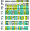
The epidemic of severe acute respiratory syndrome (SARS) was caused by a newly emerged coronavirus (SARS-CoV). Bats of several species in southern People's Republic of China harbor SARS-like CoVs and may be reservoir hosts for them. To determine whether bats in North America also harbor coronaviruses, we used reverse transcription-PCR to detect coronavirus RNA in bats. We found coronavirus RNA in 6 of 28 fecal specimens from bats of 2 of 7 species tested. The prevalence of viral RNA shedding was high: 17% in Eptesicus fuscus and 50% in Myotis occultus. Sequence analysis of a 440-bp amplicon in gene 1b showed that these Rocky Mountain bat coronaviruses formed 3 clusters in phylogenetic group 1 that were distinct from group 1 coronaviruses of Asian bats. Because of the potential for bat coronaviruses to cause disease in humans and animals, further surveillance and characterization of bat coronaviruses in North America are needed.
Figures


Similar articles
-
Genetic Characteristics of Coronaviruses from Korean Bats in 2016.Microb Ecol. 2018 Jan;75(1):174-182. doi: 10.1007/s00248-017-1033-8. Epub 2017 Jul 19. Microb Ecol. 2018. PMID: 28725945 Free PMC article.
-
Detection and phylogenetic analysis of group 1 coronaviruses in South American bats.Emerg Infect Dis. 2008 Dec;14(12):1890-3. doi: 10.3201/eid1412.080642. Emerg Infect Dis. 2008. PMID: 19046513 Free PMC article.
-
Metagenomic analysis of the viromes of three North American bat species: viral diversity among different bat species that share a common habitat.J Virol. 2010 Dec;84(24):13004-18. doi: 10.1128/JVI.01255-10. Epub 2010 Oct 6. J Virol. 2010. PMID: 20926577 Free PMC article.
-
Global Epidemiology of Bat Coronaviruses.Viruses. 2019 Feb 20;11(2):174. doi: 10.3390/v11020174. Viruses. 2019. PMID: 30791586 Free PMC article. Review.
-
Coronaviruses in Bats: A Review for the Americas.Viruses. 2021 Jun 25;13(7):1226. doi: 10.3390/v13071226. Viruses. 2021. PMID: 34201926 Free PMC article. Review.
Cited by
-
Detection of novel coronaviruses from dusky fruit bat (Penthetor lucasi) in Sarawak, Malaysian Borneo.Vet Med Sci. 2023 Nov;9(6):2634-2641. doi: 10.1002/vms3.1251. Epub 2023 Sep 2. Vet Med Sci. 2023. PMID: 37658663 Free PMC article.
-
SARS-CoV-2 wildlife surveillance in Ontario and Québec.Can Commun Dis Rep. 2022 Jun 9;48(6):243-251. doi: 10.14745/ccdr.v48i06a02. eCollection 2022 Jun 9. Can Commun Dis Rep. 2022. PMID: 37333575 Free PMC article.
-
Coronavirus sampling and surveillance in bats from 1996-2019: a systematic review and meta-analysis.Nat Microbiol. 2023 Jun;8(6):1176-1186. doi: 10.1038/s41564-023-01375-1. Epub 2023 May 25. Nat Microbiol. 2023. PMID: 37231088 Free PMC article.
-
Kathryn V. Holmes: A Career of Contributions to the Coronavirus Field.Viruses. 2022 Jul 20;14(7):1573. doi: 10.3390/v14071573. Viruses. 2022. PMID: 35891553 Free PMC article. Review.
-
Molecular, ecological, and behavioral drivers of the bat-virus relationship.iScience. 2022 Aug 19;25(8):104779. doi: 10.1016/j.isci.2022.104779. Epub 2022 Jul 20. iScience. 2022. PMID: 35875684 Free PMC article. Review.
References
Publication types
MeSH terms
Grants and funding
LinkOut - more resources
Full Text Sources
Miscellaneous

