Ancient exaptation of a CORE-SINE retroposon into a highly conserved mammalian neuronal enhancer of the proopiomelanocortin gene
- PMID: 17922573
- PMCID: PMC2000970
- DOI: 10.1371/journal.pgen.0030166
Ancient exaptation of a CORE-SINE retroposon into a highly conserved mammalian neuronal enhancer of the proopiomelanocortin gene
Abstract
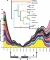
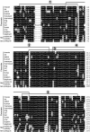
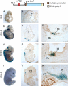
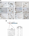
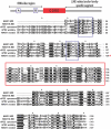
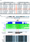
The proopiomelanocortin gene (POMC) is expressed in the pituitary gland and the ventral hypothalamus of all jawed vertebrates, producing several bioactive peptides that function as peripheral hormones or central neuropeptides, respectively. We have recently determined that mouse and human POMC expression in the hypothalamus is conferred by the action of two 5' distal and unrelated enhancers, nPE1 and nPE2. To investigate the evolutionary origin of the neuronal enhancer nPE2, we searched available vertebrate genome databases and determined that nPE2 is a highly conserved element in placentals, marsupials, and monotremes, whereas it is absent in nonmammalian vertebrates. Following an in silico paleogenomic strategy based on genome-wide searches for paralog sequences, we discovered that opossum and wallaby nPE2 sequences are highly similar to members of the superfamily of CORE-short interspersed nucleotide element (SINE) retroposons, in particular to MAR1 retroposons that are widely present in marsupial genomes. Thus, the neuronal enhancer nPE2 originated from the exaptation of a CORE-SINE retroposon in the lineage leading to mammals and remained under purifying selection in all mammalian orders for the last 170 million years. Expression studies performed in transgenic mice showed that two nonadjacent nPE2 subregions are essential to drive reporter gene expression into POMC hypothalamic neurons, providing the first functional example of an exapted enhancer derived from an ancient CORE-SINE retroposon. In addition, we found that this CORE-SINE family of retroposons is likely to still be active in American and Australian marsupial genomes and that several highly conserved exonic, intronic and intergenic sequences in the human genome originated from the exaptation of CORE-SINE retroposons. Together, our results provide clear evidence of the functional novelties that transposed elements contributed to their host genomes throughout evolution.
Conflict of interest statement
Competing interests. FSJdS, MJL, and MR have intellectual property and patent interests in the POMC neuronal-specific enhancers and have received income from the licensing of this intellectual property and related research material to financially interested companies.
Figures

Similar articles
-
Convergent evolution of two mammalian neuronal enhancers by sequential exaptation of unrelated retroposons.Proc Natl Acad Sci U S A. 2011 Sep 13;108(37):15270-5. doi: 10.1073/pnas.1104997108. Epub 2011 Aug 29. Proc Natl Acad Sci U S A. 2011. PMID: 21876128 Free PMC article.
-
The estrogen receptor α colocalizes with proopiomelanocortin in hypothalamic neurons and binds to a conserved motif present in the neuron-specific enhancer nPE2.Eur J Pharmacol. 2011 Jun 11;660(1):181-7. doi: 10.1016/j.ejphar.2010.10.114. Epub 2011 Jan 3. Eur J Pharmacol. 2011. PMID: 21211522 Free PMC article.
-
Partially redundant enhancers cooperatively maintain Mammalian pomc expression above a critical functional threshold.PLoS Genet. 2015 Feb 11;11(2):e1004935. doi: 10.1371/journal.pgen.1004935. eCollection 2015 Feb. PLoS Genet. 2015. PMID: 25671638 Free PMC article.
-
Emergence of mammals by emergency: exaptation.Genes Cells. 2010 Aug;15(8):801-12. doi: 10.1111/j.1365-2443.2010.01429.x. Epub 2010 Jul 14. Genes Cells. 2010. PMID: 20633052 Review.
-
Retroposons of salmonoid fishes (Actinopterygii: Salmonoidei) and their evolution.Gene. 2009 Apr 1;434(1-2):16-28. doi: 10.1016/j.gene.2008.04.022. Epub 2008 May 22. Gene. 2009. PMID: 18590946 Review.
Cited by
-
A mammalian tripartite enhancer cluster controls hypothalamic Pomc expression, food intake, and body weight.Proc Natl Acad Sci U S A. 2024 Apr 30;121(18):e2322692121. doi: 10.1073/pnas.2322692121. Epub 2024 Apr 23. Proc Natl Acad Sci U S A. 2024. PMID: 38652744 Free PMC article.
-
The Role of Transposable Elements of the Human Genome in Neuronal Function and Pathology.Int J Mol Sci. 2022 May 23;23(10):5847. doi: 10.3390/ijms23105847. Int J Mol Sci. 2022. PMID: 35628657 Free PMC article. Review.
-
Transposable Elements: Distribution, Polymorphism, and Climate Adaptation in Populus.Front Plant Sci. 2022 Feb 1;13:814718. doi: 10.3389/fpls.2022.814718. eCollection 2022. Front Plant Sci. 2022. PMID: 35178060 Free PMC article.
-
Retrotransposons as Drivers of Mammalian Brain Evolution.Life (Basel). 2021 Apr 22;11(5):376. doi: 10.3390/life11050376. Life (Basel). 2021. PMID: 33922141 Free PMC article. Review.
-
Genetic Mechanisms Underlying Cortical Evolution in Mammals.Front Cell Dev Biol. 2021 Feb 15;9:591017. doi: 10.3389/fcell.2021.591017. eCollection 2021. Front Cell Dev Biol. 2021. PMID: 33659245 Free PMC article. Review.
References
-
- de Souza FS, Bumaschny VF, Low MJ, Rubinstein M. Subfunctionalization of expression and peptide domains following the ancient duplication of the proopiomelanocortin gene in teleost fishes. Mol Biol Evol. 2005;22:2417–2427. - PubMed
-
- Raffin-Sanson ML, de Keyzer Y, Bertagna X. Proopiomelanocortin, a polypeptide precursor with multiple functions: from physiology to pathological conditions. Eur J Endocrinol. 2003;149:79–90. - PubMed
-
- Hadley ME, Haskell-Luevano C. The proopiomelanocortin system. Ann N Y Acad Sci. 1999;885:1–21. - PubMed
-
- Low MJ. Role of proopiomelanocortin neurons and peptides in the regulation of energy homeostasis. J Endocrinol Invest. 2004;27:95–100. - PubMed
Publication types
MeSH terms
Substances
Grants and funding
LinkOut - more resources
Full Text Sources
Other Literature Sources
Miscellaneous


