Light-powering Escherichia coli with proteorhodopsin
- PMID: 17277079
- PMCID: PMC1892948
- DOI: 10.1073/pnas.0611035104
Light-powering Escherichia coli with proteorhodopsin
Abstract
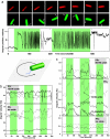
Proteorhodopsin (PR) is a light-powered proton pump identified by community sequencing of ocean samples. Previous studies have established the ecological distribution and enzymatic activity of PR, but its role in powering cells and participation in ocean energy fluxes remains unclear. Here, we show that when cellular respiration is inhibited by depleting oxygen or by the respiratory poison azide, Escherichia coli cells expressing PR become light-powered. Illumination of these cells with light coinciding with PR's absorption spectrum creates a proton motive force (pmf) that turns the flagellar motor, yielding cells that swim when illuminated with green light. By measuring the pmf of individual illuminated cells, we quantify the coupling between light-driven and respiratory proton currents, estimate the Michaelis-Menten constant (Km) of PR (10(3) photons per second/nm2), and show that light-driven pumping by PR can fully replace respiration as a cellular energy source in some environmental conditions. Moreover, sunlight-illuminated PR+ cells are less sensitive to azide than PR- cells, consistent with PR+ cells possessing an alternative means of maintaining cellular pmf and, thus, viability. Proteorhodopsin allows Escherichia coli cells to withstand environmental respiration challenges by harvesting light energy.
Conflict of interest statement
The authors declare no conflict of interest.
Figures


Similar articles
-
Proteorhodopsin is a light-driven proton pump with variable vectoriality.J Mol Biol. 2002 Aug 30;321(5):821-38. doi: 10.1016/s0022-2836(02)00696-4. J Mol Biol. 2002. PMID: 12206764
-
Proteorhodopsin Overproduction Enhances the Long-Term Viability of Escherichia coli.Appl Environ Microbiol. 2019 Dec 13;86(1):e02087-19. doi: 10.1128/AEM.02087-19. Print 2019 Dec 13. Appl Environ Microbiol. 2019. PMID: 31653788 Free PMC article.
-
Diversity and functional analysis of proteorhodopsin in marine Flavobacteria.Environ Microbiol. 2012 May;14(5):1240-8. doi: 10.1111/j.1462-2920.2012.02702.x. Epub 2012 Feb 13. Environ Microbiol. 2012. PMID: 22329552
-
[A Study on Mechanisms Underlying Proton Transport in Proton Pump-type Microbial Rhodopsins].Yakugaku Zasshi. 2023;143(2):111-118. doi: 10.1248/yakushi.22-00184. Yakugaku Zasshi. 2023. PMID: 36724923 Review. Japanese.
-
Ion-pumping microbial rhodopsins.Front Mol Biosci. 2015 Sep 22;2:52. doi: 10.3389/fmolb.2015.00052. eCollection 2015. Front Mol Biosci. 2015. PMID: 26442282 Free PMC article. Review.
Cited by
-
Scaling Transition of Active Turbulence from Two to Three Dimensions.Adv Sci (Weinh). 2024 Oct;11(38):e2402643. doi: 10.1002/advs.202402643. Epub 2024 Aug 13. Adv Sci (Weinh). 2024. PMID: 39137163 Free PMC article.
-
Engineering bionanoreactor in bacteria for efficient hydrogen production.Proc Natl Acad Sci U S A. 2024 Jul 16;121(29):e2404958121. doi: 10.1073/pnas.2404958121. Epub 2024 Jul 10. Proc Natl Acad Sci U S A. 2024. PMID: 38985767 Free PMC article.
-
Spatiotemporal dynamics of the proton motive force on single bacterial cells.Sci Adv. 2024 May 24;10(21):eadl5849. doi: 10.1126/sciadv.adl5849. Epub 2024 May 23. Sci Adv. 2024. PMID: 38781330 Free PMC article.
-
Effects of Light and Dark Conditions on the Transcriptome of Aging Cultures of Candidatus Puniceispirillum marinum IMCC1322.J Microbiol. 2024 Apr;62(4):297-314. doi: 10.1007/s12275-024-00125-0. Epub 2024 Apr 25. J Microbiol. 2024. PMID: 38662311
-
Molecular mechanisms and evolutionary robustness of a color switch in proteorhodopsins.Sci Adv. 2024 Jan 26;10(4):eadj0384. doi: 10.1126/sciadv.adj0384. Epub 2024 Jan 24. Sci Adv. 2024. PMID: 38266078 Free PMC article.
References
-
- Behrenfeld MJ, Falkowski PG. Limnol Oceanogr. 1997;42:1–20.
-
- del Giorgio PA, Duarte CM. Nature. 2002;420:379–384. - PubMed
-
- Beja O, Aravind L, Koonin EV, Suzuki MT, Hadd A, Nguyen LP, Jovanovich SB, Gates CM, Feldman RA, Spudich JL, et al. Science. 2000;289:1902–1906. - PubMed
-
- Venter JC, Remington K, Heidelberg JF, Halpern AL, Rusch D, Eisen JA, Wu D, Paulsen I, Nelson KE, Nelson W, et al. Science. 2004;304:66–74. - PubMed
Publication types
MeSH terms
Substances
LinkOut - more resources
Full Text Sources
Other Literature Sources
Research Materials


