Rewiring the severe acute respiratory syndrome coronavirus (SARS-CoV) transcription circuit: engineering a recombination-resistant genome
- PMID: 16891412
- PMCID: PMC1531645
- DOI: 10.1073/pnas.0605438103
Rewiring the severe acute respiratory syndrome coronavirus (SARS-CoV) transcription circuit: engineering a recombination-resistant genome
Abstract
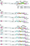
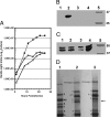
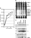
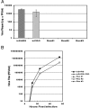
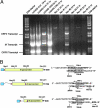
Live virus vaccines provide significant protection against many detrimental human and animal diseases, but reversion to virulence by mutation and recombination has reduced appeal. Using severe acute respiratory syndrome coronavirus as a model, we engineered a different transcription regulatory circuit and isolated recombinant viruses. The transcription network allowed for efficient expression of the viral transcripts and proteins, and the recombinant viruses replicated to WT levels. Recombinant genomes were then constructed that contained mixtures of the WT and mutant regulatory circuits, reflecting recombinant viruses that might occur in nature. Although viable viruses could readily be isolated from WT and recombinant genomes containing homogeneous transcription circuits, chimeras that contained mixed regulatory networks were invariantly lethal, because viable chimeric viruses were not isolated. Mechanistically, mixed regulatory circuits promoted inefficient subgenomic transcription from inappropriate start sites, resulting in truncated ORFs and effectively minimize viral structural protein expression. Engineering regulatory transcription circuits of intercommunicating alleles successfully introduces genetic traps into a viral genome that are lethal in RNA recombinant progeny viruses.
Conflict of interest statement
Conflict of interest statement: No conflicts declared.
Figures

Similar articles
-
Reverse genetics with a full-length infectious cDNA of severe acute respiratory syndrome coronavirus.Proc Natl Acad Sci U S A. 2003 Oct 28;100(22):12995-3000. doi: 10.1073/pnas.1735582100. Epub 2003 Oct 20. Proc Natl Acad Sci U S A. 2003. PMID: 14569023 Free PMC article.
-
Severe Acute Respiratory Syndrome (SARS) Coronavirus ORF8 Protein Is Acquired from SARS-Related Coronavirus from Greater Horseshoe Bats through Recombination.J Virol. 2015 Oct;89(20):10532-47. doi: 10.1128/JVI.01048-15. Epub 2015 Aug 12. J Virol. 2015. PMID: 26269185 Free PMC article.
-
Identification of novel subgenomic RNAs and noncanonical transcription initiation signals of severe acute respiratory syndrome coronavirus.J Virol. 2005 May;79(9):5288-95. doi: 10.1128/JVI.79.9.5288-5295.2005. J Virol. 2005. PMID: 15827143 Free PMC article.
-
Development of mouse hepatitis virus and SARS-CoV infectious cDNA constructs.Curr Top Microbiol Immunol. 2005;287:229-52. doi: 10.1007/3-540-26765-4_8. Curr Top Microbiol Immunol. 2005. PMID: 15609514 Free PMC article. Review.
-
Programmed ribosomal frameshifting in HIV-1 and the SARS-CoV.Virus Res. 2006 Jul;119(1):29-42. doi: 10.1016/j.virusres.2005.10.008. Epub 2005 Nov 28. Virus Res. 2006. PMID: 16310880 Free PMC article. Review.
Cited by
-
Molecular characteristic, evolution, and pathogenicity analysis of avian infectious bronchitis virus isolates associated with QX type in China.Poult Sci. 2024 Aug 28;103(12):104256. doi: 10.1016/j.psj.2024.104256. Online ahead of print. Poult Sci. 2024. PMID: 39288718 Free PMC article.
-
Developing Next-Generation Live Attenuated Vaccines for Porcine Epidemic Diarrhea Using Reverse Genetic Techniques.Vaccines (Basel). 2024 May 19;12(5):557. doi: 10.3390/vaccines12050557. Vaccines (Basel). 2024. PMID: 38793808 Free PMC article. Review.
-
Design and Application of Biosafe Coronavirus Engineering Systems without Virulence.Viruses. 2024 Apr 24;16(5):659. doi: 10.3390/v16050659. Viruses. 2024. PMID: 38793541 Free PMC article. Review.
-
A recombination-resistant genome for live attenuated and stable PEDV vaccines by engineering the transcriptional regulatory sequences.J Virol. 2023 Dec 21;97(12):e0119323. doi: 10.1128/jvi.01193-23. Epub 2023 Nov 16. J Virol. 2023. PMID: 37971221 Free PMC article.
-
Reverse genetics systems for SARS-CoV-2: Development and applications.Virol Sin. 2023 Dec;38(6):837-850. doi: 10.1016/j.virs.2023.10.001. Epub 2023 Oct 11. Virol Sin. 2023. PMID: 37832720 Free PMC article. Review.
References
-
- Kew O. M., Sutter R. W., de Gourville E. M., Dowdle W. R., Pallansch M. A. Annu. Rev. Microbiol. 2005;59:587–635. - PubMed
-
- Seligman S. J., Gould E. A. Lancet. 2004;363:2073–2075. - PubMed
-
- Guan Y., Zheng B. J., He Y. Q., Liu X. L., Xhuang Z. X., Cheung C. L., Luo S. W., Li P. H., Zhang L. J., Guan Y. J., et al. Science. 2003;302:276–278. - PubMed
-
- Li W., Shi Z., Yu M., Ren W., Smith C., Epstein J. H., Wang H., Crameri G., Hu Z., Zhang H., et al. Science. 2005;310:676–679. - PubMed
Publication types
MeSH terms
Grants and funding
LinkOut - more resources
Full Text Sources
Other Literature Sources
Miscellaneous


