Energy transduction: proton transfer through the respiratory complexes
- PMID: 16756489
- PMCID: PMC2659341
- DOI: 10.1146/annurev.biochem.75.062003.101730
Energy transduction: proton transfer through the respiratory complexes
Abstract
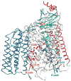
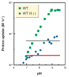
A series of metalloprotein complexes embedded in a mitochondrial or bacterial membrane utilize electron transfer reactions to pump protons across the membrane and create an electrochemical potential (DeltamuH+). Current understanding of the principles of electron-driven proton transfer is discussed, mainly with respect to the wealth of knowledge available from studies of cytochrome c oxidase. Structural, experimental, and theoretical evidence supports the model of long-distance proton transfer via hydrogen-bonded water chains in proteins as well as the basic concept that proton uptake and release in a redox-driven pump are driven by charge changes at the membrane-embedded centers. Key elements in the pumping mechanism may include bound water, carboxylates, and the heme propionates, arginines, and associated water above the hemes. There is evidence for an important role of subunit III and proton backflow, but the number and nature of gating mechanisms remain elusive, as does the mechanism of physiological control of efficiency.
Figures


Similar articles
-
An arginine to lysine mutation in the vicinity of the heme propionates affects the redox potentials of the hemes and associated electron and proton transfer in cytochrome c oxidase.Biochemistry. 2005 Aug 9;44(31):10457-65. doi: 10.1021/bi050283d. Biochemistry. 2005. PMID: 16060654 Free PMC article.
-
Water chain formation and possible proton pumping routes in Rhodobacter sphaeroides cytochrome c oxidase: a molecular dynamics comparison of the wild type and R481K mutant.Biochemistry. 2005 Aug 9;44(31):10475-85. doi: 10.1021/bi0502902. Biochemistry. 2005. PMID: 16060656
-
Proton-coupled electron transfer drives the proton pump of cytochrome c oxidase.Nature. 2006 Apr 6;440(7085):829-32. doi: 10.1038/nature04619. Nature. 2006. PMID: 16598262
-
Cytochrome c oxidase: chemistry of a molecular machine.Adv Enzymol Relat Areas Mol Biol. 1995;71:79-208. doi: 10.1002/9780470123171.ch3. Adv Enzymol Relat Areas Mol Biol. 1995. PMID: 8644492 Review.
-
Respiratory conservation of energy with dioxygen: cytochrome C oxidase.Met Ions Life Sci. 2015;15:89-130. doi: 10.1007/978-3-319-12415-5_4. Met Ions Life Sci. 2015. PMID: 25707467 Review.
Cited by
-
Electron transfer in the respiratory chain at low salinity.Nat Commun. 2024 Sep 19;15(1):8241. doi: 10.1038/s41467-024-52475-3. Nat Commun. 2024. PMID: 39300056 Free PMC article.
-
Circadian coupling of mitochondria in a deep-diving mammal.J Exp Biol. 2024 Apr 1;227(7):jeb246990. doi: 10.1242/jeb.246990. Epub 2024 Apr 8. J Exp Biol. 2024. PMID: 38495024 Free PMC article.
-
Proton transfer reactions: From photochemistry to biochemistry and bioenergetics.BBA Adv. 2023 Mar 9;3:100085. doi: 10.1016/j.bbadva.2023.100085. eCollection 2023. BBA Adv. 2023. PMID: 37378355 Free PMC article.
-
Preventative effect of TSPO ligands on mixed antibody-mediated rejection through a Mitochondria-mediated metabolic disorder.J Transl Med. 2023 May 2;21(1):295. doi: 10.1186/s12967-023-04134-2. J Transl Med. 2023. PMID: 37131248 Free PMC article.
-
High-Throughput Screening and Molecular Dynamics Simulation of Natural Products for the Identification of Anticancer Agents against MCM7 Protein.Appl Bionics Biomech. 2022 Sep 15;2022:8308192. doi: 10.1155/2022/8308192. eCollection 2022. Appl Bionics Biomech. 2022. Retraction in: Appl Bionics Biomech. 2023 Dec 20;2023:9828706. doi: 10.1155/2023/9828706 PMID: 36157125 Free PMC article. Retracted.
References
-
- Mills DA, Schmidt B, Hiser C, Westley E, Ferguson-Miller S. J Biol Chem. 2002;277:14894–901. - PubMed
-
- Kadenbach B. Biochim Biophys Acta. 2003;1604:77–94. - PubMed
-
- Brand MD, Buckingham JA, Esteves TC, Green K, Lambert AJ, et al. Biochem Soc Symp. 2004;71:203–13. - PubMed
-
- Lanyi JK. Annu Rev Physiol. 2004;66:665–88. - PubMed
-
- Hunte C, Palsdottir H, Trumpower BL. FEBS Lett. 2003;545:39–46. - PubMed
Publication types
MeSH terms
Substances
Grants and funding
LinkOut - more resources
Full Text Sources


