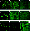
Serotype differences and lack of biofilm formation characterize Yersinia pseudotuberculosis infection of the Xenopsylla cheopis flea vector of Yersinia pestis
- PMID: 16428415
- PMCID: PMC1347331
- DOI: 10.1128/JB.188.3.1113-1119.2006
Serotype differences and lack of biofilm formation characterize Yersinia pseudotuberculosis infection of the Xenopsylla cheopis flea vector of Yersinia pestis
Abstract
Yersinia pestis, the agent of plague, is usually transmitted by fleas. To produce a transmissible infection, Y. pestis colonizes the flea midgut and forms a biofilm in the proventricular valve, which blocks normal blood feeding. The enteropathogen Yersinia pseudotuberculosis, from which Y. pestis recently evolved, is not transmitted by fleas. However, both Y. pestis and Y. pseudotuberculosis form biofilms that adhere to the external mouthparts and block feeding of Caenorhabditis elegans nematodes, which has been proposed as a model of Y. pestis-flea interactions. We compared the ability of Y. pestis and Y. pseudotuberculosis to infect the rat flea Xenopsylla cheopis and to produce biofilms in the flea and in vitro. Five of 18 Y. pseudotuberculosis strains, encompassing seven serotypes, including all three serotype O3 strains tested, were unable to stably colonize the flea midgut. The other strains persisted in the flea midgut for 4 weeks but did not increase in numbers, and none of the 18 strains colonized the proventriculus or produced a biofilm in the flea. Y. pseudotuberculosis strains also varied greatly in their ability to produce biofilms in vitro, but there was no correlation between biofilm phenotype in vitro or on the surface of C. elegans and the ability to colonize or block fleas. Our results support a model in which a genetic change in the Y. pseudotuberculosis progenitor of Y. pestis extended its pre-existing ex vivo biofilm-forming ability to the flea gut environment, thus enabling proventricular blockage and efficient flea-borne transmission.
Figures


Similar articles
-
Identification of gmhA, a Yersinia pestis gene required for flea blockage, by using a Caenorhabditis elegans biofilm system.Infect Immun. 2005 Nov;73(11):7236-42. doi: 10.1128/IAI.73.11.7236-7242.2005. Infect Immun. 2005. PMID: 16239518 Free PMC article.
-
Comparative Ability of Oropsylla montana and Xenopsylla cheopis Fleas to Transmit Yersinia pestis by Two Different Mechanisms.PLoS Negl Trop Dis. 2017 Jan 12;11(1):e0005276. doi: 10.1371/journal.pntd.0005276. eCollection 2017 Jan. PLoS Negl Trop Dis. 2017. PMID: 28081130 Free PMC article.
-
Acute oral toxicity of Yersinia pseudotuberculosis to fleas: implications for the evolution of vector-borne transmission of plague.Cell Microbiol. 2007 Nov;9(11):2658-66. doi: 10.1111/j.1462-5822.2007.00986.x. Epub 2007 Jun 24. Cell Microbiol. 2007. PMID: 17587333
-
The evolution of flea-borne transmission in Yersinia pestis.Curr Issues Mol Biol. 2005 Jul;7(2):197-212. Curr Issues Mol Biol. 2005. PMID: 16053250 Review.
-
Yersinia pestis biofilm in the flea vector and its role in the transmission of plague.Curr Top Microbiol Immunol. 2008;322:229-48. doi: 10.1007/978-3-540-75418-3_11. Curr Top Microbiol Immunol. 2008. PMID: 18453279 Free PMC article. Review.
Cited by
-
Yersinia pestis Δail Mutants Are Not Susceptible to Human Complement Bactericidal Activity in the Flea.Appl Environ Microbiol. 2023 Feb 28;89(2):e0124422. doi: 10.1128/aem.01244-22. Epub 2023 Feb 6. Appl Environ Microbiol. 2023. PMID: 36744930 Free PMC article.
-
Cpx-signalling facilitates Hms-dependent biofilm formation by Yersinia pseudotuberculosis.NPJ Biofilms Microbiomes. 2022 Mar 29;8(1):13. doi: 10.1038/s41522-022-00281-4. NPJ Biofilms Microbiomes. 2022. PMID: 35351893 Free PMC article.
-
Molecular and Genetic Mechanisms That Mediate Transmission of Yersinia pestis by Fleas.Biomolecules. 2021 Feb 3;11(2):210. doi: 10.3390/biom11020210. Biomolecules. 2021. PMID: 33546271 Free PMC article. Review.
-
A Trimeric Autotransporter Enhances Biofilm Cohesiveness in Yersinia pseudotuberculosis but Not in Yersinia pestis.J Bacteriol. 2020 Sep 23;202(20):e00176-20. doi: 10.1128/JB.00176-20. Print 2020 Sep 23. J Bacteriol. 2020. PMID: 32778558 Free PMC article.
-
Genomic Epidemiology and Phenotyping Reveal on-Farm Persistence and Cold Adaptation of Raw Milk Outbreak-Associated Yersinia pseudotuberculosis.Front Microbiol. 2019 May 14;10:1049. doi: 10.3389/fmicb.2019.01049. eCollection 2019. Front Microbiol. 2019. PMID: 31156582 Free PMC article.
References
-
- Beloin, C., and J. M. Ghigo. 2005. Finding gene-expression patterns in bacterial biofilms. Trends Microbiol. 13:16-19. - PubMed
-
- Blanc, G., and M. Balthazard. 1944. Contribution a l'etude du comportement de microbes pathogenes chez la puce du rat Xenopyslla cheopis. Le bacille de la pseudo-tuberculose des rongeurs. C. R. Soc. Biol. Paris 138:811-812.
Publication types
MeSH terms
Grants and funding
LinkOut - more resources
Full Text Sources


