iBet uBet web content aggregator. Adding the entire web to your favor.


Link to original content: https://pubmed.ncbi.nlm.nih.gov/11466280
Transduction by phiBB-1, a bacteriophage of Borrelia burgdorferi - PubMed Skip to main page content
U.S. flag

An official website of the United States government

Dot gov

The .gov means it’s official.
Federal government websites often end in .gov or .mil. Before sharing sensitive information, make sure you’re on a federal government site.

Https

The site is secure.
The https:// ensures that you are connecting to the official website and that any information you provide is encrypted and transmitted securely.

Access keys NCBI Homepage MyNCBI Homepage Main Content Main Navigation
. 2001 Aug;183(16):4771-8.
doi: 10.1128/JB.183.16.4771-4778.2001.

Transduction by phiBB-1, a bacteriophage of Borrelia burgdorferi

Affiliations

Transduction by phiBB-1, a bacteriophage of Borrelia burgdorferi

C H Eggers et al. J Bacteriol. 2001 Aug.

Abstract

We previously described a bacteriophage of the Lyme disease agent Borrelia burgdorferi designated phiBB-1. This phage packages the host complement of the 32-kb circular plasmids (cp32s), a group of homologous molecules found throughout the genus Borrelia. To demonstrate the ability of phiBB-1 to package and transduce DNA, a kanamycin resistance cassette was inserted into a cloned fragment of phage DNA, and the resulting construct was transformed into B. burgdorferi CA-11.2A cells. The kan cassette recombined into a resident cp32 and was stably maintained. The cp32 containing the kan cassette was packaged by phiBB-1 released from this B. burgdorferi strain. phiBB-1 has been used to transduce this antibiotic resistance marker into naive CA-11.2A cells, as well as two other strains of B. burgdorferi. This is the first direct evidence of a mechanism for lateral gene transfer in B. burgdorferi.


Figures
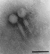
FIG. 1
FIG. 1
B. burgdorferi phage particles. Samples were collected from polyethylene glycol-precipitated cell supernatants of an MNNG-treated culture of B. burgdorferi CA-11.2A and viewed by transmission electron microscopy. Although previously reported as having a simple noncontractile tail (12), subsequent modifications to the purification and preparation protocol (10, 11) reveal that all phage particles observed have intact contractile sheaths, seen here either extended (left) or contracted (right). Phosphotungstic acid stain. Bar, 45 nm.
FIG. 2
FIG. 2
Kanamycin-resistant transformants of B. burgdorferi. (A) pCE210 construct. A 4-kb fragment of phage DNA (solid line) was cloned into the HindIII site of pBluescript SK+ (dashed line). The kan cassette (hatched box) was cloned into a unique NdeI site on the phage fragment 1.1 kb from the SK primer location. The locations of the SK, KS, cp32SK12NdeIF (cp32F), cp32SK12NdeIR (cp32R), and KanR1207F (kanF) oligonucleotides are shown (not drawn to scale). Arrows indicate oligonucleotide site and orientation. (B) Transformed CA-11.2A colonies were screened by PCR using the cp32SK12NdeI primers. PCR products were resolved on a 1% agarose gel and stained with EtBr. Both a small product (no kan cassette; black arrow) and a larger product (integrated kan cassette; hatched arrow) were amplified from the transformants. (C) Products consistent with a population of cp32s both containing the kan cassette (hatched arrow) and lacking the kan cassette (black arrow) were also amplified from phage DNA collected from CA-11.2A/kanR transformants. pCE210 DNA was amplified as a positive (+) control, and wild-type CA-11.2A phage DNA was used as a negative (−) control.
FIG. 3
FIG. 3
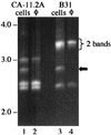
Determining the minimum number of cp32 molecules in B. burgdorferi cells and φBB-1 capsids by PCR amplification of VR1. Highly conserved primers that flank a variable region (VR1) of cp32 amplify four different size products from B. burgdorferi strains CA-11.2A and B31. The two VR1s from the cp32s of B31 at ∼3.3 kb can be resolved with extended electrophoresis times (see Fig. 7). PCR of φBB-1 released from both of these strains amplifies three of the VR1 products (lanes 2 and 4), but not the ∼2.8-kb amplicon (black arrow; see Table 2). Fragments were resolved on a 0.8% agarose gel by FIGE and stained with EtBr. Molecular sizes (in kilobase pairs) are indicated.
FIG. 4
FIG. 4
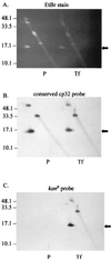
Analysis of the genomic location of the kanamycin resistance cassette. Total DNA from both parental CA-11.2A (P) and a transformed clone of CA-11.2A/kanR (Tf) was extracted and resolved by two-dimensional gel electrophoresis. The gel was stained with EtBr (A), blotted to nylon, and probed with the cp32-specific probe 4 (B) or pOK12, the source of the kan gene (C). The kan probe has the same hybridization pattern as the cp32-specific probe. The supercoiled form of cp32 is indicated by the black arrow, while the other hybridization sites likely represent the nicked (or lp56) and linearized forms of cp32 generated during DNA isolation. Molecular sizes (in kilobase pairs) are indicated.
FIG. 5
FIG. 5
Analysis of kan cassette packaging by φBB-1. Phage particles were precipitated from both parental (P) and transformed (Tf) CA-11.2A cells. The DNA was extracted and resolved on a 0.5% agarose gel by conventional field electrophoresis. The gel was stained with EtBr (A), then blotted and probed with either probe 4 (cp32-specific; B) or pOK12 (kan; C). Molecular sizes (in kilobase pairs) are indicated.
FIG. 6
FIG. 6
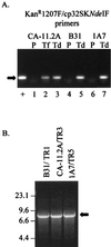
PCR analysis of kanamycin-resistant transductants of B. burgdorferi. (A) Putative transductants from B. burgdorferi strains CA-11.2A, B31, and 1A7 were screened by PCR using the KanR1207F and cp32SK12NdeIF primers. pCE210 was amplified as a positive control (+), while the parental cells (P) of each strain served as negative controls (lanes 1, 4, and 6). Lane 2 is the CA-11.2A/kanR transformant (Tf). PCR yields a ∼125-bp product from DNA containing the kan cassette (black arrow, lanes 2, 3, 5, and 7). The products were resolved on a 1% agarose gel and stained by EtBr. (B) The kan integration site in each transductant was verified by long-range PCR using the KanR1207F and cp32-VR1R primers.
FIG. 7
FIG. 7
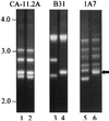
Amplification of variable region VR1 from B. burgdorferi kan transductants. The cp32-VR1 primers were used to amplify the cp32 VR1s of parental CA-11.2A (lane 1), B31 (lane 3), and 1A7 (lane 5) as well as transductants CA-11.2A/TR3 (lane 2), B31/TR1 (lane 4), and 1A7/TR5 (lane 6). The 2,537-bp VR1 (black arrow) was found in all transductants. Amplification products were resolved on 0.8% agarose gels by FIGE and stained with EtBr. Molecular sizes are shown in kilobase pairs.
FIG. 8
FIG. 8
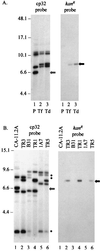
Restriction mapping of the cp32s of B. burgdorferi kan transductants. (A) Comparison of the restriction maps of parental, transformant, and transductant CA-11.2A DNA. Plasmid DNA was extracted from CA-11.2A (lane 1; P), the CA-11.2A/kanR transformant (lane 2; Tf), and CA-11.2A/TR3 (lane 3; Td), digested with EcoRV, and resolved by FIGE. A blot of the gel was probed either with the cp32SK12NdeI PCR product (left panel) or with kan (right panel). A unique ∼6.6-kb fragment of CA-11.2A parental DNA is indicated by the hatched arrow. A ∼8-kb band (black arrow) corresponding to the ∼6.6-kb band plus the 1.3-kb kan insert is present in both the transformant and the transductant. (B) Comparison of the restriction maps of the cp32s of B. burgdorferi kan transductants. Plasmid DNA from CA-11.2A (lane 1), CA-11.2A/TR3 (lane 2), B31 (lane 3), B31/TR1 (lane 4), 1A7 (lane 5), and 1A7/TR5 (lane 6) was digested with XbaI and resolved by FIGE. A blot of the gel was probed with either the cp32SK12NdeI probe (left panel) or the kan probe (right panel). A ∼8-kb band (black arrow) is found only in the transductants. The hatched arrow indicates the 6.6-kb fragment from the parental CA-11.2A strain (lacking the kan cassette). Three bands (∗) appear in parental B31 but not B31/TR1. Molecular sizes are shown in kilobase pairs.

Similar articles

Cited by

References

    1. Akins D R, Caimano M J, Yang X, Cerna F, Norgard M V, Radolf J D. Molecular and evolutionary analysis of Borrelia burgdorferi 297 circular plasmid-encoded lipoproteins with OspE- and OspF-like leader peptides. Infect Immun. 1999;67:1526–1532. - PMC - PubMed
    1. Ausubel F M, Brent R, Kingston R E, Moore D D, Seidman J G, Smith J A, Struhl K, editors. Short protocols in molecular biology. New York, N.Y: John Wiley & Sons, Inc.; 1999.
    1. Barbour A G, Hayes S F. Biology of Borrelia species. Microbiol Rev. 1986;50:381–400. - PMC - PubMed
    1. Bono J L, Elias A F, Kupko J J, Stevenson B, Tilly K, Rosa P. Efficient targeted mutagenesis in Borrelia burgdorferi. J Bacteriol. 2000;182:2445–2452. - PMC - PubMed
    1. Burgdorfer W, Barbour A G, Hayes S F, Benach J L, Grunwaldt E, Davis J P. Lyme disease–a tick-borne spirochetosis? Science. 1982;216:1317–1319. - PubMed

Publication types

LinkOut - more resources