iBet uBet web content aggregator. Adding the entire web to your favor.


Link to original content: https://pubmed.ncbi.nlm.nih.gov/10373536
Fanconi anemia proteins FANCA, FANCC, and FANCG/XRCC9 interact in a functional nuclear complex - PubMed Skip to main page content
U.S. flag

An official website of the United States government

Dot gov

The .gov means it’s official.
Federal government websites often end in .gov or .mil. Before sharing sensitive information, make sure you’re on a federal government site.

Https

The site is secure.
The https:// ensures that you are connecting to the official website and that any information you provide is encrypted and transmitted securely.

Access keys NCBI Homepage MyNCBI Homepage Main Content Main Navigation
. 1999 Jul;19(7):4866-73.
doi: 10.1128/MCB.19.7.4866.

Fanconi anemia proteins FANCA, FANCC, and FANCG/XRCC9 interact in a functional nuclear complex

Affiliations

Fanconi anemia proteins FANCA, FANCC, and FANCG/XRCC9 interact in a functional nuclear complex

I Garcia-Higuera et al. Mol Cell Biol. 1999 Jul.

Abstract

Fanconi anemia (FA) is an autosomal recessive cancer susceptibility syndrome with at least eight complementation groups (A to H). Three FA genes, corresponding to complementation groups A, C, and G, have been cloned, but their cellular function remains unknown. We have previously demonstrated that the FANCA and FANCC proteins interact and form a nuclear complex in normal cells, suggesting that the proteins cooperate in a nuclear function. In this report, we demonstrate that the recently cloned FANCG/XRCC9 protein is required for binding of the FANCA and FANCC proteins. Moreover, the FANCG protein is a component of a nuclear protein complex containing FANCA and FANCC. The amino-terminal region of the FANCA protein is required for FANCG binding, FANCC binding, nuclear localization, and functional activity of the complex. Our results demonstrate that the three cloned FA proteins cooperate in a large multisubunit complex. Disruption of this complex results in the specific cellular and clinical phenotype common to most FA complementation groups.


Figures

FIG. 1
FIG. 1
Characterization of the MMC sensitivities of FA-A and FA-G lymphoblast lines. The full-length FANCG cDNA (7, 25) was isolated by RT-PCR of RNA from a human MG63 tumor cell line and subcloned into the murine retroviral vector pMMP(puro) (19). The indicated retroviral supernatants were generated and used to infect FA lymphoblast lines, and puromycin-resistant cells were selected. The cells analyzed included EUFA316 (FA-G) cells, HSC72 (FA-A) cells, and normal (PD7) cells. Consistent with previous studies (7), pMMP-FANCG infection also resulted in functional complementation (correction of MMC sensitivity) of another FA-G cell line, EUFA143 (data not shown). WT, wild type.
FIG. 2
FIG. 2
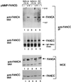
Expression of FANCG in FA-G lymphoblasts restores the FANCA/FANCC interaction. Whole-cell extracts (WCE) were generated from lymphoblast lines, including EUFA316, EUFA316-FANCG, EUFA143, EUFA143-FANCG, and PD7 (normal adult control). These protein extracts (100 μg) were probed directly by immunoblotting with either anti-FANCA or anti-FANCC serum. The FANCA protein is indicated by an arrow, and additional bands in the anti-FANCA immunoblot are nonspecific. Alternatively, the same amount of protein from each extract (2 mg) was used for immunoprecipitation with affinity-purified anti-FANCC serum as indicated. Immune complexes were resolved by sodium dodecyl sulfate-polyacrylamide gel electrophoresis, transferred to nitrocellulose, and immunoblotted with anti-FANCA or anti-FANCC serum. WT, wild type; IP, immunoprecipitation. The values to the right of the gel are molecular sizes in kilodaltons.
FIG. 3
FIG. 3
FANCG is a component of the cytoplasmic and nuclear FANCA/FANCC protein complex. (A) The indicated lymphoblast lines, including EUFA316 (FA-G cells), EUFA316 corrected with the FANCG cDNA (FA-G + FANCG), or PD7 (normal adult control) were lysed and fractionated into cytoplasmic and nuclear fractions. Proteins from each fraction were immunoprecipitated with control nonimmune (P), anti-FANCA (A), anti-FANCG (G), or anti-FANCC (C) serum. Proteins were resolved by sodium dodecyl sulfate-polyacrylamide gel electrophoresis, transferred to nitrocellulose, and immunoblotted with anti-FANCA (upper panel), anti-FANCG (middle panel) or anti-FANCC (lower panel) serum. To demonstrate that the absence of the FANCA/FANCC interaction in FA-G cells is not simply due to the low expression level of FANCA and FANCC in these cells, three times more cytoplasmic extract (6 mg in 1 ml) was used for EUFA316 (lanes 1 to 4) than for the other two cell lines (lanes 5 to 12). (B) To ensure effective fractionation, samples from the indicated lymphoblast lines described in panel A were analyzed for topoisomerase levels (upper panel) and β-tubulin levels (lower panel). WT, wild type; IgG, immunoglobulin G; IP, immunoprecipitating.
FIG. 4
FIG. 4
Binding of FANCA and FANCG in vitro. In vitro-translated, [35S]methionine-labeled FANCA (A), FANCG (G), and FANCC (C) were prepared in separate reactions and mixed as indicated (Input Protein). Immunoprecipitation was done with either preimmune (P), anti-FANCA (A), anti-FANCG (G), or anti-FANCC (C) serum. Total reticulocyte lysate (without immunoprecipitation) containing labeled FANCA, FANCG, and FANCC was loaded in lanes 13, 14, and 15, respectively. IP, immunoprecipitating.
FIG. 5
FIG. 5
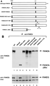
The amino-terminal NLS region of FANCA is required for FANCG binding and functional activity. (A) Schematic representation of wild-type (WT) FANCA and mutant proteins. The wild-type FANCA protein is 1,455 amino acids long and contains a bipartite NLS and a partial leucine zipper (LZ) motif. The FANCA-ΔNLS mutant protein is missing the amino-terminal 35 amino acids. The FANCA-NLS-mut2 protein has all of the basic amino acids of the NLS mutated. The SV40T-FANCA protein contains the 13-amino-acid NLS of the SV40 T antigen in place of the NLS of FANCA. The FANCA(H1110P) and FANCA(R1117G) mutations are based on FANCA mutational screens (22). cDNAs encoding these mutant FANCA proteins were generated and expressed in the FA-A fibroblast line GM6914 as previously described (29). (B) Cell lysates were prepared from GM6914 FA-A fibroblasts expressing the indicated FANCA mutant proteins. The FANCA proteins were immunoprecipitated with an anti-FANCA serum (anti-carboxy-terminal antibody), and the immune complexes were analyzed by anti-FANCA and anti-FANCG immunoblotting. As a negative control, GM6914 cells expressing nlsLacZ (no FANCA protein) were analyzed (lane 2). For the FANCA-ΔXho mutant protein, immunoprecipitation was done with the anti-FANCA (anti-amino-terminal) antibody as previously described (29). IP, immunoprecipitation.
FIG. 6
FIG. 6
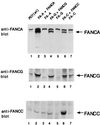
Analysis of the FANCA/FANCG/FANCC protein complex in cell lines from multiple FA complementation groups. (A) Whole-cell extracts were prepared from the indicated lymphoblast lines, and proteins were immunoprecipitated with either preimmune (P) or anti-FANCA (A) serum. Immune complexes were immunoblotted with antiserum to FANCA, FANCG, or FANCC, as indicated. The cell lines analyzed included normal control PD7 cells (lanes 1 and 2) and HSC72 (lanes 3 and 4), HSC72–FA-A (lanes 5 and 6), BD32 (lanes 7 and 8), HSC230 (lanes 9 and 10), PD4 (lanes 11 and 12), HSC536 (lanes 13 and 14), PD20L (lanes 15 and 16), EUFA130 (lanes 17 and 18), EUFA121 (lanes 19 and 20), EUFA316 (lanes 21 and 22), and EUFA173 (lanes 23 and 24) cells. The BD32 (FA-A) cell line expressed a mutant FANCA polypeptide [FANCA(H1110P)]. The HSC536 (FA-C) cell line expressed a mutant FANCC polypeptide [FANCC(L554P)]. All of the cell lines examined expressed comparatively equal levels of FANCC protein as judged by whole-cell lysate immunoblotting (data not shown), except PD4 cells, which expressed no FANCC protein. IP, immunoprecipitating; IgG, immunoglobulin G. WT, wild type.
FIG. 7
FIG. 7
The FANCA, FANCG, and FANCC proteins stabilize the FA protein complex. Whole-cell extracts from the indicated lymphoblast lines were prepared, and the same amount of protein from each was resolved by sodium dodecyl sulfate-polyacrylamide gel electrophoresis, transferred to nitrocellulose, and immunoblotted with affinity-purified antiserum against the FANCA, FANCC, or FANCG protein, as indicated. WT, wild type.
FIG. 8
FIG. 8
Nuclear localization of the FANCA and FANCG polypeptides. The human fibroblast line GM0637, derived from a normal (wild-type) control, was infected with either pMMP-FANCA (wild type) (A) or pMMP-FANCG (wild type) (B) as indicated. Pools of infected cells were stained with anti-FANCA or anti-FANCG serum or stained with the DNA-specific dye 4′,6-diamidino-2-phenylindole (DAPI) and analyzed by immunofluorescence assay as previously described (29).
FIG. 9
FIG. 9
Schematic model of molecular interactions of the FANCA, FANCG, and FANCC proteins. Based on available data, at least two regions of the FANCA polypeptide are required for the FANCA-FANCG-FANCC interaction. First, the amino-terminal region of the FANCA protein, which includes the bipartite NLS region, is required for interaction with the FANCG protein. Second, a region near the carboxy terminus of FANCA is required for the FANCC interaction. Patient-derived point mutations in this region disrupt the FANCC interaction. The FANCC interaction is a weak or regulated interaction, requiring FANCA/FANCG binding and the products of other FA genes (44). The region of FANCG required for FANCA binding is not known. The region of FANCC required for FANCA binding may be the carboxy terminus of FANCC. This region is highly conserved among human, murine, and bovine FANCC proteins. Also, an FA-C patient-derived point mutation [FANCC(L554P)] ablates FANCA binding (21).

Similar articles

Cited by

References

    1. Auerbach A D, Buchwald M, Joenje H. Fanconi anemia. In: Vogelstein B, Kinzler K W, editors. The genetic basis of human cancer. New York, N.Y: McGraw Hill; 1997.
    1. Baumann P, West S C. Role of the human RAD51 protein in homologous recombination and double-stranded break repair. Trends Biochem Sci. 1998;23:247–251. - PubMed
    1. Buchwald M. Complementation groups: one or more per gene. Nat Genet. 1995;11:228–230. - PubMed
    1. Chu G. Double strand break repair. J Biol Chem. 1997;272:24097–24100. - PubMed
    1. Cumming R C, Liu J M, Youssoufian H, Buchwald M. Suppression of apoptosis in hematopoietic factor-dependent progenitor cell lines by expression of the FAC gene. Blood. 1996;88:4558–4567. - PubMed

Publication types

Substances

LinkOut - more resources