All-trans and 9-cis retinoic acid alter rat hepatic stellate cell phenotype differentially
- PMID: 10369717
- PMCID: PMC1727592
- DOI: 10.1136/gut.45.1.134
All-trans and 9-cis retinoic acid alter rat hepatic stellate cell phenotype differentially
Abstract
Background: Hepatic stellate cells exert specific functions in the liver: storage of large amounts of retinyl esters, synthesis and breakdown of hepatic extracellular matrix, secretion of a variety of cytokines, and control of the diameter of the sinusoids.
Aims: To examine the influence of all-trans retinoic acid (ATRA) and 9-cis retinoic acid (9RA) on extracellular matrix production and proliferation of activated hepatic stellate cells.
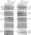
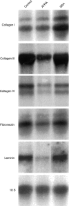
Methods: Cells were isolated using collagenase/pronase, purified by centrifugation in nycodenz, and cultured for two weeks. At this time point the cells exhibited the activated phenotype. Cells were exposed to various concentrations of ATRA and 9RA. The expression of procollagens I, III, and IV, of fibronectin and of laminin were analysed by immunoprecipitation and northern hybridisation.
Results: ATRA exerted a significant inhibitory effect on the synthesis of procollagens type I, III, and IV, fibronectin, and laminin, but did not influence stellate cell proliferation, whereas 9RA showed a clear but late effect on proliferation. 9RA increased procollagen I mRNA 1.9-fold, but did not affect the expression of other matrix proteins.
Conclusion: Results showed that ATRA and 9RA exert different, often contrary effects on activated stellate cells. These observations may explain prior divergent results obtained following retinoid administration to cultured stellate cells or in animals subjected to fibrogenic stimuli.
Figures


Similar articles
-
Differential modulation of rat hepatic stellate phenotype by natural and synthetic retinoids.Hepatology. 2004 Jan;39(1):97-108. doi: 10.1002/hep.20015. Hepatology. 2004. PMID: 14752828
-
Vitamin A inhibits pancreatic stellate cell activation: implications for treatment of pancreatic fibrosis.Gut. 2006 Jan;55(1):79-89. doi: 10.1136/gut.2005.064543. Epub 2005 Jul 25. Gut. 2006. PMID: 16043492 Free PMC article.
-
Effect of tumour necrosis factor-alpha on proliferation, activation and protein synthesis of rat hepatic stellate cells.J Hepatol. 1997 Dec;27(6):1067-80. doi: 10.1016/s0168-8278(97)80151-1. J Hepatol. 1997. PMID: 9453433
-
Prevention of cultured rat stellate cell transformation and endothelin-B receptor upregulation by retinoic acid.Br J Pharmacol. 2003 Jun;139(4):765-74. doi: 10.1038/sj.bjp.0705303. Br J Pharmacol. 2003. PMID: 12813000 Free PMC article.
-
Beauty is in the eye of the beholder: emerging concepts and pitfalls in hepatic stellate cell research.J Hepatol. 2002 Oct;37(4):527-35. doi: 10.1016/s0168-8278(02)00263-5. J Hepatol. 2002. PMID: 12217608 Review. No abstract available.
Cited by
-
Resistance to Gemcitabine in Pancreatic Ductal Adenocarcinoma: A Physiopathologic and Pharmacologic Review.Cancers (Basel). 2022 May 18;14(10):2486. doi: 10.3390/cancers14102486. Cancers (Basel). 2022. PMID: 35626089 Free PMC article. Review.
-
Association of metabolic dysfunction-associated fatty liver disease with kidney disease.Nat Rev Nephrol. 2022 Apr;18(4):259-268. doi: 10.1038/s41581-021-00519-y. Epub 2022 Jan 10. Nat Rev Nephrol. 2022. PMID: 35013596 Review.
-
Cellular and Molecular Mechanisms Underlying Liver Fibrosis Regression.Cells. 2021 Oct 15;10(10):2759. doi: 10.3390/cells10102759. Cells. 2021. PMID: 34685739 Free PMC article. Review.
-
Cellular Mechanisms of Liver Fibrosis.Front Pharmacol. 2021 May 6;12:671640. doi: 10.3389/fphar.2021.671640. eCollection 2021. Front Pharmacol. 2021. PMID: 34025430 Free PMC article. Review.
-
PNPLA3-A Potential Therapeutic Target for Personalized Treatment of Chronic Liver Disease.Front Med (Lausanne). 2019 Dec 17;6:304. doi: 10.3389/fmed.2019.00304. eCollection 2019. Front Med (Lausanne). 2019. PMID: 31921875 Free PMC article. Review.
References
Publication types
MeSH terms
Substances
LinkOut - more resources
Full Text Sources

