iBet uBet web content aggregator. Adding the entire web to your favor.


Link to original content: http://www.rcsb.org/structure/5G4M
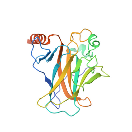
RCSB PDB - 5G4M: Crystal structure of the p53 cancer mutant Y220C in complex with a monofluorinated derivative of the small molecule stabilizer Phikan083

 5G4M

Crystal structure of the p53 cancer mutant Y220C in complex with a monofluorinated derivative of the small molecule stabilizer Phikan083


Experimental Data Snapshot

  • Method: X-RAY DIFFRACTION
  • Resolution: 1.38 Å
  • R-Value Free: 0.170 
  • R-Value Work: 0.146 
  • R-Value Observed: 0.147 

Starting Model: experimental
View more details

wwPDB Validation   3D Report Full Report


Ligand Structure Quality Assessment 


This is version 1.4 of the entry. See complete history


Literature

Harnessing Fluorine-Sulfur Contacts and Multipolar Interactions for the Design of P53 Mutant Y220C Rescue Drugs.

Bauer, M.R.Jones, R.N.Baud, M.G.J.Wilcken, R.Boeckler, F.M.Fersht, A.R.Joerger, A.C.Spencer, J.

(2016) ACS Chem Biol 11: 2265

  • DOI: https://doi.org/10.1021/acschembio.6b00315
  • Primary Citation of Related Structures:  
    5G4M, 5G4N, 5G4O

  • PubMed Abstract: 

    Many oncogenic mutants of the tumor suppressor p53 are conformationally unstable, including the frequently occurring Y220C mutant. We have previously developed several small-molecule stabilizers of this mutant. One of these molecules, PhiKan083, 1-(9-ethyl-9H-carbazole-3-yl)-N-methylmethanamine, binds to a mutation-induced surface crevice with a KD = 150 μM, thereby increasing the melting temperature of the protein and slowing its rate of aggregation. Incorporation of fluorine atoms into small molecule ligands can substantially improve binding affinity to their protein targets. We have, therefore, harnessed fluorine-protein interactions to improve the affinity of this ligand. Step-wise introduction of fluorines at the carbazole ethyl anchor, which is deeply buried within the binding site in the Y220C-PhiKan083 complex, led to a 5-fold increase in affinity for a 2,2,2-trifluoroethyl anchor (ligand efficiency of 0.3 kcal mol(-1) atom(-1)). High-resolution crystal structures of the Y220C-ligand complexes combined with quantum chemical calculations revealed favorable interactions of the fluorines with protein backbone carbonyl groups (Leu145 and Trp146) and the sulfur of Cys220 at the mutation site. Affinity gains were, however, only achieved upon trifluorination, despite favorable interactions of the mono- and difluorinated anchors with the binding pocket, indicating a trade-off between energetically favorable protein-fluorine interactions and increased desolvation penalties. Taken together, the optimized carbazole scaffold provides a promising starting point for the development of high-affinity ligands to reactivate the tumor suppressor function of the p53 mutant Y220C in cancer cells.


  • Organizational Affiliation

    MRC Laboratory of Molecular Biology , Francis Crick Avenue, Cambridge Biomedical Campus, Cambridge CB2 0QH, United Kingdom.


Macromolecules
Find similar proteins by:  (by identity cutoff)  |  3D Structure
Entity ID: 1
MoleculeChains Sequence LengthOrganismDetailsImage
CELLULAR TUMOR ANTIGEN P53
A, B
219Homo sapiensMutation(s): 5 
UniProt & NIH Common Fund Data Resources
Find proteins for P04637 (Homo sapiens)
Explore P04637 
Go to UniProtKB:  P04637
PHAROS:  P04637
GTEx:  ENSG00000141510 
Entity Groups  
Sequence Clusters30% Identity50% Identity70% Identity90% Identity95% Identity100% Identity
UniProt GroupP04637
Sequence Annotations
Expand
  • Reference Sequence
Experimental Data & Validation

Experimental Data

  • Method: X-RAY DIFFRACTION
  • Resolution: 1.38 Å
  • R-Value Free: 0.170 
  • R-Value Work: 0.146 
  • R-Value Observed: 0.147 
  • Space Group: P 21 21 21
Unit Cell:
Length ( Å )Angle ( ˚ )
a = 65.03α = 90
b = 71.144β = 90
c = 105.144γ = 90
Software Package:
Software NamePurpose
PHENIXrefinement
PHENIXphasing

Structure Validation

View Full Validation Report


Ligand Structure Quality Assessment 


Entry History 

Deposition Data

Revision History  (Full details and data files)

  • Version 1.0: 2016-06-22
    Type: Initial release
  • Version 1.1: 2016-09-28
    Changes: Database references
  • Version 1.2: 2019-05-08
    Changes: Data collection, Experimental preparation, Other
  • Version 1.3: 2019-05-15
    Changes: Data collection, Experimental preparation
  • Version 1.4: 2024-01-10
    Changes: Data collection, Database references, Derived calculations, Other, Refinement description