iBet uBet web content aggregator. Adding the entire web to your favor.


Link to original content: http://www.rcsb.org/pdb/explore.do?structureId=8CR8
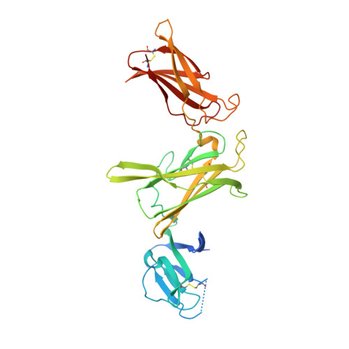
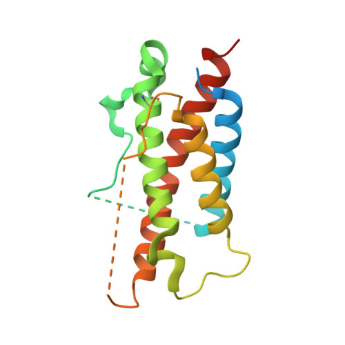
RCSB PDB - 8CR8: human Interleukin-23

 8CR8

human Interleukin-23


Experimental Data Snapshot

  • Method: X-RAY DIFFRACTION
  • Resolution: 2.00 Å
  • R-Value Free: 0.256 
  • R-Value Work: 0.235 
  • R-Value Observed: 0.236 

Starting Model: experimental
View more details

wwPDB Validation   3D Report Full Report


This is version 1.2 of the entry. See complete history


Literature

Structures of complete extracellular receptor assemblies mediated by IL-12 and IL-23.

Bloch, Y.Felix, J.Merceron, R.Provost, M.Symakani, R.A.De Backer, R.Lambert, E.Mehdipour, A.R.Savvides, S.N.

(2024) Nat Struct Mol Biol 31: 591-597

  • DOI: https://doi.org/10.1038/s41594-023-01190-6
  • Primary Citation of Related Structures:  
    8C7M, 8CR5, 8CR6, 8CR8, 8ODX, 8ODZ, 8OE0, 8OE4, 8PB1, 8PPM

  • PubMed Abstract: 

    Cell-surface receptor complexes mediated by pro-inflammatory interleukin (IL)-12 and IL-23, both validated therapeutic targets, are incompletely understood due to the lack of structural insights into their complete extracellular assemblies. Furthermore, there is a paucity of structural details describing the IL-12-receptor interaction interfaces, in contrast to IL-23-receptor complexes. Here we report structures of fully assembled mouse IL-12/human IL-23-receptor complexes comprising the complete extracellular segments of the cognate receptors determined by electron cryo-microscopy. The structures reveal key commonalities but also surprisingly diverse features. Most notably, whereas IL-12 and IL-23 both utilize a conspicuously presented aromatic residue on their α-subunit as a hotspot to interact with the N-terminal Ig domain of their high-affinity receptors, only IL-12 juxtaposes receptor domains proximal to the cell membrane. Collectively, our findings will help to complete our understanding of cytokine-mediated assemblies of tall cytokine receptors and will enable a cytokine-specific interrogation of IL-12/IL-23 signaling in physiology and disease.


  • Organizational Affiliation

    Unit for Structural Biology, Department of Biochemistry and Microbiology, Ghent University, Ghent, Belgium.


Macromolecules
Find similar proteins by:  (by identity cutoff)  |  3D Structure
Entity ID: 1
MoleculeChains Sequence LengthOrganismDetailsImage
Interleukin-12 subunit beta325Homo sapiensMutation(s): 1 
Gene Names: IL12BNKSF2
UniProt & NIH Common Fund Data Resources
Find proteins for P29460 (Homo sapiens)
Explore P29460 
Go to UniProtKB:  P29460
PHAROS:  P29460
GTEx:  ENSG00000113302 
Entity Groups  
Sequence Clusters30% Identity50% Identity70% Identity90% Identity95% Identity100% Identity
UniProt GroupP29460
Glycosylation
Glycosylation Sites: 1Go to GlyGen: P29460-1
Sequence Annotations
Expand
  • Reference Sequence
Find similar proteins by:  (by identity cutoff)  |  3D Structure
Entity ID: 2
MoleculeChains Sequence LengthOrganismDetailsImage
Interleukin-23 subunit alpha196Homo sapiensMutation(s): 0 
Gene Names: IL23ASGRFUNQ2498/PRO5798
UniProt & NIH Common Fund Data Resources
Find proteins for Q9NPF7 (Homo sapiens)
Explore Q9NPF7 
Go to UniProtKB:  Q9NPF7
PHAROS:  Q9NPF7
GTEx:  ENSG00000110944 
Entity Groups  
Sequence Clusters30% Identity50% Identity70% Identity90% Identity95% Identity100% Identity
UniProt GroupQ9NPF7
Sequence Annotations
Expand
  • Reference Sequence
Oligosaccharides

Help

Entity ID: 3
MoleculeChains Length2D Diagram Glycosylation3D Interactions
alpha-D-mannopyranose-(1-2)-alpha-D-mannopyranose-(1-6)-[alpha-D-mannopyranose-(1-3)]beta-D-mannopyranose-(1-4)-2-acetamido-2-deoxy-beta-D-glucopyranose-(1-4)-2-acetamido-2-deoxy-beta-D-glucopyranose
C
6N-Glycosylation
Glycosylation Resources
GlyTouCan:  G60415BS
GlyCosmos:  G60415BS
GlyGen:  G60415BS
Small Molecules
Ligands 1 Unique
IDChains Name / Formula / InChI Key2D Diagram3D Interactions
TB
Query on TB

Download Ideal Coordinates CCD File 
D [auth A]TERBIUM(III) ION
Tb
HKCRVXUAKWXBLE-UHFFFAOYSA-N
Experimental Data & Validation

Experimental Data

  • Method: X-RAY DIFFRACTION
  • Resolution: 2.00 Å
  • R-Value Free: 0.256 
  • R-Value Work: 0.235 
  • R-Value Observed: 0.236 
  • Space Group: P 1 21 1
Unit Cell:
Length ( Å )Angle ( ˚ )
a = 67.748α = 90
b = 59.842β = 98.06
c = 69.989γ = 90
Software Package:
Software NamePurpose
PHENIXrefinement
XDSdata reduction
XDSdata scaling
PHASERphasing

Structure Validation

View Full Validation Report


Entry History & Funding Information

Deposition Data


Funding OrganizationLocationGrant Number
Research Foundation - Flanders (FWO)Belgium12S0519N

Revision History  (Full details and data files)

  • Version 1.0: 2024-02-07
    Type: Initial release
  • Version 1.1: 2024-05-01
    Changes: Database references
  • Version 1.2: 2024-11-06
    Changes: Structure summary