Rotavirus as an Expression Platform of Domains of the SARS-CoV-2 Spike Protein
- PMID: 34063562
- PMCID: PMC8147602
- DOI: 10.3390/vaccines9050449
Rotavirus as an Expression Platform of Domains of the SARS-CoV-2 Spike Protein
Abstract
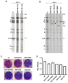
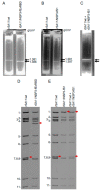
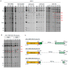
Among vaccines administered to children are those targeting rotavirus, a segmented double-stranded RNA virus that represents a major cause of severe gastroenteritis. To explore the feasibility of establishing a combined rotavirus-SARS-CoV-2 vaccine, we generated recombinant (r)SA11 rotaviruses with modified segment 7 RNAs that contained coding cassettes for NSP3, a translational 2A stop-restart signal, and a FLAG-tagged portion of the SARS-CoV-2 spike (S) protein: S1 fragment, N-terminal domain (NTD), receptor-binding domain (RBD), extended RBD (ExRBD), or S2 core (CR) domain. Generation of rSA11 containing the S1 coding sequence required a sequence insertion of 2.2 kbp, the largest such insertion yet introduced into the rotavirus genome. Immunoblotting showed that rSA11 viruses containing the smaller NTD, RBD, ExRBD, and CR coding sequences expressed S-protein products of expected size, with ExRBD expressed at highest levels. These rSA11 viruses were genetically stable during serial passage. In contrast, the rSA11 virus containing the full-length S coding sequence (rSA11/NSP3-fS1) failed to express its expected 80 kDa fS1 product, for unexplained reasons. Moreover, rSA11/NSP3-fS1 was genetically unstable, with variants lacking the S1 insertion appearing during serial passage. Nonetheless, these results emphasize the potential usefulness of rotavirus vaccines as expression vectors of immunogenic portions of the SARS-CoV-2 S protein, including NTD, RBD, ExRBD, and CR, that have sizes smaller than the S1 fragment.
Keywords: COVID-19 vaccine; Reoviridae; SARS-CoV-2; expression vector; reverse genetics; rotavirus; rotavirus vaccine; spike protein.
Conflict of interest statement
J.T.P. is an inventor on a patent application related to the content of this paper.
Figures


Update of
-
Rotavirus as an Expression Platform of the SARS-CoV-2 Spike Protein.bioRxiv [Preprint]. 2021 Feb 18:2021.02.18.431835. doi: 10.1101/2021.02.18.431835. bioRxiv. 2021. Update in: Vaccines (Basel). 2021 May 03;9(5):449. doi: 10.3390/vaccines9050449 PMID: 33619485 Free PMC article. Updated. Preprint.
Similar articles
-
Recombinant rotavirus expressing the glycosylated S1 protein of SARS-CoV-2.J Gen Virol. 2023 Oct;104(10):001899. doi: 10.1099/jgv.0.001899. J Gen Virol. 2023. PMID: 37830788 Free PMC article.
-
Rotavirus as an Expression Platform of the SARS-CoV-2 Spike Protein.bioRxiv [Preprint]. 2021 Feb 18:2021.02.18.431835. doi: 10.1101/2021.02.18.431835. bioRxiv. 2021. Update in: Vaccines (Basel). 2021 May 03;9(5):449. doi: 10.3390/vaccines9050449 PMID: 33619485 Free PMC article. Updated. Preprint.
-
Generation of Recombinant Rotavirus Expressing NSP3-UnaG Fusion Protein by a Simplified Reverse Genetics System.J Virol. 2019 Nov 26;93(24):e01616-19. doi: 10.1128/JVI.01616-19. Print 2019 Dec 15. J Virol. 2019. PMID: 31597761 Free PMC article.
-
Emergence, evolution, and vaccine production approaches of SARS-CoV-2 virus: Benefits of getting vaccinated and common questions.Saudi J Biol Sci. 2022 Apr;29(4):1981-1997. doi: 10.1016/j.sjbs.2021.12.020. Epub 2021 Dec 13. Saudi J Biol Sci. 2022. PMID: 34924802 Free PMC article. Review.
-
SARS-CoV-2 Spike Protein Extrapolation for COVID Diagnosis and Vaccine Development.Front Mol Biosci. 2021 Jul 28;8:607886. doi: 10.3389/fmolb.2021.607886. eCollection 2021. Front Mol Biosci. 2021. PMID: 34395515 Free PMC article. Review.
Cited by
-
VP4 Mutation Boosts Replication of Recombinant Human/Simian Rotavirus in Cell Culture.Viruses. 2024 Apr 5;16(4):565. doi: 10.3390/v16040565. Viruses. 2024. PMID: 38675907 Free PMC article.
-
Production of OSU G5P[7] Porcine Rotavirus Expressing a Fluorescent Reporter via Reverse Genetics.Viruses. 2024 Mar 7;16(3):411. doi: 10.3390/v16030411. Viruses. 2024. PMID: 38543776 Free PMC article.
-
Recovery of Recombinant Rotaviruses by Reverse Genetics.Methods Mol Biol. 2024;2733:249-263. doi: 10.1007/978-1-0716-3533-9_15. Methods Mol Biol. 2024. PMID: 38064037
-
A Novel Rotavirus Reverse Genetics Platform Supports Flexible Insertion of Exogenous Genes and Enables Rapid Development of a High-Throughput Neutralization Assay.Viruses. 2023 Sep 30;15(10):2034. doi: 10.3390/v15102034. Viruses. 2023. PMID: 37896813 Free PMC article.
-
Recombinant rotavirus expressing the glycosylated S1 protein of SARS-CoV-2.J Gen Virol. 2023 Oct;104(10):001899. doi: 10.1099/jgv.0.001899. J Gen Virol. 2023. PMID: 37830788 Free PMC article.
References
-
- Pollán M., Pérez-Gómez B., Pastor-Barriuso R., Oteo J., Hernán M.A., Pérez-Olmeda M., Sanmartín J.L., Fernández-García A., Cruz I., Fernández de Larrea N., et al. Prevalence of SARS-CoV-2 in Spain (ENE-COVID): A nationwide, population-based seroepidemiological study. Lancet. 2020;396:535–544. doi: 10.1016/S0140-6736(20)31483-5. - DOI - PMC - PubMed
Grants and funding
LinkOut - more resources
Full Text Sources
Other Literature Sources
Miscellaneous


