iBet uBet web content aggregator. Adding the entire web to your favor.


Link to original content: http://www.ncbi.nlm.nih.gov/pubmed/28875267
Kingdom Chromista and its eight phyla: a new synthesis emphasising periplastid protein targeting, cytoskeletal and periplastid evolution, and ancient divergences - PubMed Skip to main page content
U.S. flag

An official website of the United States government

Dot gov

The .gov means it’s official.
Federal government websites often end in .gov or .mil. Before sharing sensitive information, make sure you’re on a federal government site.

Https

The site is secure.
The https:// ensures that you are connecting to the official website and that any information you provide is encrypted and transmitted securely.

Access keys NCBI Homepage MyNCBI Homepage Main Content Main Navigation
. 2018 Jan;255(1):297-357.
doi: 10.1007/s00709-017-1147-3. Epub 2017 Sep 5.

Kingdom Chromista and its eight phyla: a new synthesis emphasising periplastid protein targeting, cytoskeletal and periplastid evolution, and ancient divergences

Affiliations

Kingdom Chromista and its eight phyla: a new synthesis emphasising periplastid protein targeting, cytoskeletal and periplastid evolution, and ancient divergences

Thomas Cavalier-Smith. Protoplasma. 2018 Jan.

Abstract

In 1981 I established kingdom Chromista, distinguished from Plantae because of its more complex chloroplast-associated membrane topology and rigid tubular multipartite ciliary hairs. Plantae originated by converting a cyanobacterium to chloroplasts with Toc/Tic translocons; most evolved cell walls early, thereby losing phagotrophy. Chromists originated by enslaving a phagocytosed red alga, surrounding plastids by two extra membranes, placing them within the endomembrane system, necessitating novel protein import machineries. Early chromists retained phagotrophy, remaining naked and repeatedly reverted to heterotrophy by losing chloroplasts. Therefore, Chromista include secondary phagoheterotrophs (notably ciliates, many dinoflagellates, Opalozoa, Rhizaria, heliozoans) or walled osmotrophs (Pseudofungi, Labyrinthulea), formerly considered protozoa or fungi respectively, plus endoparasites (e.g. Sporozoa) and all chromophyte algae (other dinoflagellates, chromeroids, ochrophytes, haptophytes, cryptophytes). I discuss their origin, evolutionary diversification, and reasons for making chromists one kingdom despite highly divergent cytoskeletons and trophic modes, including improved explanations for periplastid/chloroplast protein targeting, derlin evolution, and ciliary/cytoskeletal diversification. I conjecture that transit-peptide-receptor-mediated 'endocytosis' from periplastid membranes generates periplastid vesicles that fuse with the arguably derlin-translocon-containing periplastid reticulum (putative red algal trans-Golgi network homologue; present in all chromophytes except dinoflagellates). I explain chromist origin from ancestral corticates and neokaryotes, reappraising tertiary symbiogenesis; a chromist cytoskeletal synapomorphy, a bypassing microtubule band dextral to both centrioles, favoured multiple axopodial origins. I revise chromist higher classification by transferring rhizarian subphylum Endomyxa from Cercozoa to Retaria; establishing retarian subphylum Ectoreta for Foraminifera plus Radiozoa, apicomonad subclasses, new dinozoan classes Myzodinea (grouping Colpovora gen. n., Psammosa), Endodinea, Sulcodinea, and subclass Karlodinia; and ranking heterokont Gyrista as phylum not superphylum.

Keywords: Chloroplast protein targeting; Chromist evolution; Chromist periplastid membrane; Chromist periplastid reticulum; Microtubular centriolar roots; Sporozoan conoid origin.


Conflict of interest statement

The author declares he has no conflict of interest.

Figures
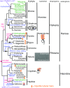
Fig. 1
Fig. 1
Relationships between major chromist groups inferred from sequence trees mostly using many scores of genes. For taxa ranked as subphyla or lower, clades still possessing the ancestral chromist plastid of red algal origin are shown in green, and purely heterotrophic ones without evidence for plastids are shown in black. Black discs mark inferred extremely early plastid losses. Too little is known about protalveolates, bigyromonads, and heterotrophic Hacrobia to know whether they retain DNA-free colourless plastids like most heterotrophic Dinozoa or not. Paraphyletic bigyromonads (mostly still uncultured) are not broken down into constituent clades. Major harosan innovations discussed here are shown in blue; for the detailed treatment of hacrobian cell diversification, see Cavalier-Smith et al. (2015a). The best nuclear, plastid, and mitochondrial trees all show this topology (see text); though topologically accurate, this diagram is temporally extremely misleading: branch lengths do not represent time. Virtually, all bifurcations shown occurred in the Precambrian >600 My ago; the basal stems occupied only a tiny fraction of the ~ 750 My history of Chromista (Cavalier-Smith et al. ; Cavalier-Smith, in prep.). Two lateral gene transfers (LGTs) from bacteria (purple) prove that ancestral Myzozoa and Hacrobia each had plastids and effectively eliminate the possibility that ochrophytes could have arisen from either of them by a late tertiary symbiogenesis (lateral plastid transfer). The LGT into the ancestral hacrobian plastid is especially important as showing that plastids were present immediately after the very first chromist bifurcation. Ancestral chromists were haploid biciliates with younger anterior cilium (blue) and older posterior cilium (black, typically with different structures and beat patterns produced by ciliary transformation in its second cell cycle). Ciliates (Ciliophora) multiplied cilia in kineties and evolved separate somatic multiploid macronuclei (Ma) and diploid germline micronuclei (Mi) and complex mouths to make giant multiciliate cells, whereas some chromists lost cilia altogether, exemplified by the micrograph of an endomyxan rhizarian Filoreta (Bass et al. 2009a, b) that evolved a remarkable net-like multinucleate body. Nucleomorphs (NMs) were lost twice independently in photosynthetic lineages (phycobilins lost simultaneously) and additionally in all heterotrophs but Chilomonas
Fig. 2
Fig. 2
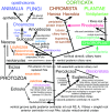
Schematic eukaryote phylogeny fully consistent with 187-protein trees (Cavalier-Smith et al. 2015a), rooted as in a 72-protein archaebacteria-rooted ribosomal tree (Raymann et al.’s Fig. 1), showing relations amongst the five eukaryote kingdoms (upper case). Kingdom Chromista comprising subkingdoms Harosa (Heterokonta, Alveolata, and infrakingdom Rhizaria) and Hacrobia (phyla Haptista and Cryptista) is most closely related to Plantae that consists of three major groups with distinct chloroplast pigments and ultrastructure: Glaucophyta and Rhodophyta (both with phycobilisomes, unstacked thylakoids, and cytosolic starch) and Viridiplantae with chlorophyll b instead of phycobilisomes, stacked thylakoids, and plastid starch. Plant chloroplasts evolved by a single primary enslavement of a cyanobacterium with both phycobilisomes and chlorophyll a (green arrow) and chromist plastids evolved by a single secondary symbiogenetic enslavement of a red alga (red arrow). All seven phyla of basal kingdom Protozoa are shown, subdivided into two subkingdoms, Neozoa and Eozoa. The four neozoan phyla (Choanozoa, Amoebozoa, Sulcozoa, Loukozoa) are more closely related to animals and Fungi than to superkingdom Corticata (Plantae plus Chromista) or to Eozoa: collectively animals, fungi, and Neozoa are an entirely non-photosynthetic clade (scotokaryotes: Cavalier-Smith et al. 2015a). Scotokaryotes are sisters of corticates if the tree is correctly rooted, forming joint clade neokaryotes. Eozoa being a clade sister to neokaryotes (He et al. 2014) or within neokaryotes (Derelle et al. 2015) rather than ancestral as shown is cell biologically improbable. Phyla Eolouka and Percolozoa have the most primitive mitochondrial genomes (Kamikawa et al. 2014) and retain ancestral bacterial cytochrome c biogenesis unlike derived neokaryotes and Euglenozoa (Cavalier-Smith 2010). Irrespective of the precise position of the eukaryote root, excavate protozoa (orange; defined as ancestrally biciliates having posterior ciliary vane and ventral feeding groove with an homologous microtubular/fibrillar cytoskeleton of three distinctive posterior centriolar roots (Simpson 2003), but no cortical alveoli; contrary to past usages, excavates here exclude the cytoskeletally radically different discicristates as well as Tsukubomonas with the simplest cytoskeleton of all biciliate Eozoa) are paraphyletic ancestors of Sulcozoa (which arose by evolving a dorsal pellicle and posterior ciliary gliding: Cavalier-Smith ; Cavalier-Smith et al. 2014) and Corticata, which arose by evolving cortical alveoli and simple ciliary hairs whilst originally retaining all neoloukan cytoskeletal microtubular roots—all evident in the harosan alveolate subphylum Protalveolata whose orders Colponemida and Acavomonadida still feed by directing prey into the groove by a vaned posterior cilium exactly as in the neoloukan excavate Malawimonas (phylum Neolouka here includes secondarily anaerobic subphylum Metamonada: Cavalier-Smith ; Cavalier-Smith et al. 2015a). As the text explains, the ancestors of chromists almost certainly used this groove-based feeding before they evolved BB and tubular ciliary hairs and enslaved red algal plastids. Orthokaryotes (named here for the putative clade comprising neokaryotes and cytoskeletally distinct Jakobea, i.e. excavates sensu stricto plus all their descendants) ancestrally had two orthogonal centrioles (parallel in discicristates except Pharyngomonas), orthodox stacked Golgi (arguably ancestrally unstacked in Tsukubamonas and Percolozoa), two opposite posterior ciliary roots (Tsukubamonas only one, its singlet root inherently part of R2), and always orthodox nuclear gene transcriptional control that evolved in the ancestral eukaryote (lost by Euglenozoa)
Fig. 3
Fig. 3
Contrasting membrane topology of Plantae and algal Chromista (superkingdom Corticata). Plantae (a) originated by primary enslavement of a cyanobacterium to make plastids and Chromista (b, c) by secondary intracellular enslavement of a red algal plant. Both target nuclear-coded proteins to plastids by transit peptides (TPs) recognised by outer membrane (OM, blue) Toc receptors and to mitochondria (enslaved α-proteobacteria) by topogenic sequences recognised by OM Tom receptors. For clarity, Golgi shown only in c and peroxisomes and lysosomes omitted. a Cyanophora, from the earliest diverging plant phylum Glaucophyta. Plastid membrane topology is identical to cyanobacteria with thylakoids. The common ancestor of red algae and green plants (not shown) lost cortical alveoli (which grow by fusion of Golgi-derived vesicles), red algae and two green plant subgroups lost chloroplast envelope murein peptidoglycan, and green plants lost phycobilisomes and stack their thylakoids. b Cryptophytes retain the enslaved red algal nucleus (simplified to a tiny nucleomorph), starch, and cytosolic ribosomes within the periplastid space (PS), and phycobilins (shown in red but can be blue instead) in the thylakoid lumen; all other euchromists (haptophytes, Ochrophytina, not shown) lost these four components and stack their thylakoids in threes not pairs, but like cryptophytes retained the red algal plasma membrane as the periplastid membrane (PPM) and a periplastid reticulum (PR) here argued to be the relict trans-Golgi network (TGN) of the enslaved red alga and topologically distinct from the PPM. c Myzozoa lack periplastid ribosomes, phycobilins, and nucleomorph DNA; thylakoids are stacked in threes; PPM (present in Apicomplexa—red dashed line; lost in Dinozoa) and plastid are not within the rough ER. The original phagosome membrane (now epiplastid membrane, EpM) remains smooth and receives vesicles (V) containing nucleus-encoded plastid proteins from the Golgi. Dinozoa lack PR, but Apicomplexa have a likely homologue (not shown)
Fig. 4
Fig. 4
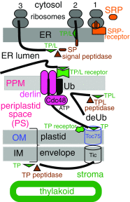
The standard model for import of nuclear-coded proteins into the chromist periplastid space (PS) and plastids, ignoring a possible role for the periplastid reticulum (PR). Ribosomes are shown in successive stages of protein synthesis and translocation (13). Nascent imported proteins (thick black line) have an N-terminal signal peptide (SP, brown oblong) that projects from the large ribosomal subunit and is recognised by a signal recognition particle (SRP) that then binds to an ER SRP receptor, ensuring that the ribosome attaches to the ER and extrudes the whole protein through an ER-embedded Sec14 channel (not shown) into the ER lumen. An ER lumenal signal peptidase cleaves off SP, exposing subterminal TP/TPL (green triangle) which is recognised by still unidentified dual purpose TP/TPL receptors (green) on the PPM and transferred to a membrane-embedded derlin oligomer essential for transfer to the PS (this stage only is more complex if PR is involved: Fig. 5). Derlin-mediated translocation into the PS depends on preprotein ubiquitinylation by ubiquitinating enzymes (Ub within PPM plus PS cofactors) and a PS-specific ubiquitin (pUb). pUb-tagged proteins are recognised by a PS-located Cdc48 ATPase, which (helped by cofactors) actively pulls them into the PS where ubiquitin is removed by deubiquitinating enzymes (deUb). Proteins that function within PS (e.g. Cdc48, deubiquitinating enzymes, TPL peptidase in all chromists, starch-making enzymes in cryptophytes, proteasomal proteins in cryptophytes, and heterokonts) have their TPL removed by a TPL peptidase, as must nuclear-coded NM proteins like DNA polymerases in cryptophytes. Imported proteins with a TP rather than TPL pass onwards into the plastid stroma through the standard Toc/Tic plastid envelope outer membrane (OM) and inner membrane (IM) channels, TP being removed later in the stroma by a different TP peptidase. Nuclear-coded intrathylakoid proteins often have tripartite N-terminal topogenic sequences with a second SP downstream of TP for transport across the thylakoid membrane using stromal insertion machinery
Fig. 5
Fig. 5
Possible role for periplastid reticulum (PR) in protein import to the chromist periplastid space (PS) and plastid. The essential difference from the standard model (Fig. 4) is that for preprotein extrusion into the PS, mature oligomeric derlin (A in cryptophytes, B in other chromists) and its associated ubiquitinating enzyme (Ub) are postulated to function not in the PPM (Fig. 4) but in the PR membrane where their assembly into the functional derlin/Ub macromolecular complex is postulated to occur. Individual derlin As and Ub are NM coded and made on periplastid ribosomes in cryptophytes and can enter PR by direct insertion from the PS. In other euchromists and Apicomplexa, derlin and Ub are nuclear coded (two distinct derlin B paralogues in heterokonts and haptophytes; one in apicomplexa) and postulated to be carried individually (like all other proteins with bipartite targeting sequences) from the ER lumen by TP/L receptor-mediated endocytic PPM budding (stage 4) to form periplastid endocytic vesicles (PEV) that fuse with PR (stage 5) to place PS- and plastid-destined preproteins into the PR lumen. TP/TPL-labelled preproteins put into the PR lumen by PEV fusion are extruded into the PS via the derlin/Ub complex powered by Cdc48 ATP hydrolysis (stage 6) and further processed (stages 8–9) exactly as in the standard model (Fig. 4). Separate PS transport vesicles (PRVs) are assumed to bud from the PR and fuse with the PPM, recycling TP/L receptors and lipids to the PPM (stage 7), including any lipids newly synthesised by the PR. Preprotein entry into the space outside the PPM differs in euchromists (cotranslational: upper left) and myzozoan alveolates [posttranslational: upper right: via fusion (d) of vesicles (CV) with the epiplastid membrane (EpM) that arose by TP/L receptor-mediated budding (c) from the Golgi, put there by vesicles budded (b) from the rough ER where preproteins entered its lumen cotranslationally (a)]. Dinozoa secondarily simplified plastid preprotein import by losing PPM, PR, and separate PS proteins, attaching EpM directly to OM
Fig. 6
Fig. 6
Cytoskeletal innovations during corticate and chromist origins. Left diagrams summarise the ancestral condition in excavate ancestors of corticates as represented by the loukozoan Malawimonas. Upper shows the whole cell seen from the right with the feeding groove tilted obliquely to show left and right mt roots (R1, R2) that support feeding groove rims and floor. Younger anterior cilium (C2) with oar-like beat and older posterior cilium (C1) undulating from base to tip simultaneously propel the cell forward (arrow) and waft food into the groove for ingestion. Lower left (Loukozoa) and right (ancestral Chromista) diagrams view the cell apex from the ventral side (so the cell’s right is on the left) to show mt arrays (colour: mt bands R1R3; plus a dorsal fan of diverging mts that support the cell’s dorsal surface) and associated fibrous supports (black: AC, I). The orthogonal centrioles (anterior A, posterior P) are interconnected by asymmetric linkers and in Loukozoa (left) a dorsal mt fan and anterior left mt band (R3) connect C2s to the apical dorsal plasma membrane. R3 is developmental precursor of R1. The ancestral corticate interposed novel cortical alveoli between the plasma membrane and dorsal fan, which split into a right bypassing mt band (BB) and numerous single, diverging subpellicular mts attached to alveolar inner faces. Chromists (right) initially kept all these cytoskeletal components, modifying them as centrioles moved subapically as the text explains. Their sister Plantae lost BB, the R2 outer branch, and B fibres. A second anterior right root R4 (not shown; see text) evolved polyphyletically by heterochrony in several chromist and plant lineages as a simplified developmental precursor of R2 (1 or few mts). The text argues that developmentally and evolutionarily the singlet root (S, brown) is a specialised R2 subcomponent, not a third posterior root as traditionally assumed. Dorsal fan and apical mts are actually longitudinal (as shown for BB only); the purple line symbolises a cross section of their mt arrays

Similar articles

Cited by

References

    1. Agrawal S, et al. An apicoplast localized ubiquitylation system is required for the import of nuclear-encoded plastid proteins. PLoS Pathog. 2013;9:e1003426. doi: 10.1371/journal.ppat.1003426. - DOI - PMC - PubMed
    1. Agrawal S, van Dooren GG, Beatty WL, Striepen B. Genetic evidence that an endosymbiont-derived endoplasmic reticulum-associated protein degradation (ERAD) system functions in import of apicoplast proteins. J Biol Chem. 2009;284:33683–33691. doi: 10.1074/jbc.M109.044024. - DOI - PMC - PubMed
    1. Aleoshin VV, Mylnikov AP, Mirzaeva GS, Mikhailov KV, Karpov SA. Heterokont predator Develorapax marinus gen. et sp. nov.—a model of the ochrophyte ancestor. Frontiers Microbiol. 2016;7:1194. doi: 10.3389/fmicb.2016.01194. - DOI - PMC - PubMed
    1. Amiar S, et al. Apicoplast-localized lysophosphatidic acid precursor assembly is required for bulk phospholipid synthesis in Toxoplasma gondii and relies on an algal/plant-like glycerol 3-phosphate acyltransferase. PLoS Pathog. 2016;12:e1005765. doi: 10.1371/journal.ppat.1005765. - DOI - PMC - PubMed
    1. Anderson OR, Cavalier-Smith T. Ultrastructure of Diplophrys parva, a new small freshwater species, and a revised analysis of Labyrinthulea (Heterokonta) Acta Protozool. 2012;51:291–304.

LinkOut - more resources