A sunblock based on bioadhesive nanoparticles
- PMID: 26413985
- PMCID: PMC4654636
- DOI: 10.1038/nmat4422
A sunblock based on bioadhesive nanoparticles
Abstract
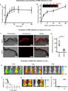
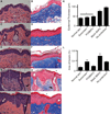
The majority of commercial sunblock preparations use organic or inorganic ultraviolet (UV) filters. Despite protecting against cutaneous phototoxicity, direct cellular exposure to UV filters has raised a variety of health concerns. Here, we show that the encapsulation of padimate O (PO)--a model UV filter--in bioadhesive nanoparticles (BNPs) prevents epidermal cellular exposure to UV filters while enhancing UV protection. BNPs are readily suspended in water, facilitate adherence to the stratum corneum without subsequent intra-epidermal or follicular penetration, and their interaction with skin is water resistant yet the particles can be removed via active towel drying. Although the sunblock based on BNPs contained less than 5 wt% of the UV-filter concentration found in commercial standards, the anti-UV effect was comparable when tested in two murine models. Moreover, the BNP-based sunblock significantly reduced double-stranded DNA breaks when compared with a commercial sunscreen formulation.
Figures


Similar articles
-
Benefit and risk of organic ultraviolet filters.Regul Toxicol Pharmacol. 2001 Jun;33(3):285-99. doi: 10.1006/rtph.2001.1476. Regul Toxicol Pharmacol. 2001. PMID: 11407932 Review.
-
Photostability and efficacy studies of topical formulations containing UV-filters combination and vitamins A, C and E.Int J Pharm. 2007 Oct 1;343(1-2):181-9. doi: 10.1016/j.ijpharm.2007.05.048. Epub 2007 May 26. Int J Pharm. 2007. PMID: 17614223
-
Biodegradable bioadhesive nanoparticle incorporation of broad-spectrum organic sunscreen agents.Bioeng Transl Med. 2018 Jul 6;4(1):129-140. doi: 10.1002/btm2.10092. eCollection 2019 Jan. Bioeng Transl Med. 2018. PMID: 30680324 Free PMC article.
-
There is no influence of a temperature rise on in vivo adsorption of UV filters into the stratum corneum.J Dermatol Sci. 2001 Oct;27(2):77-81. doi: 10.1016/s0923-1811(01)00100-1. J Dermatol Sci. 2001. PMID: 11532370
-
Nanocarriers and Microcarriers for Enhancing the UV Protection of Sunscreens: An Overview.J Pharm Sci. 2019 Dec;108(12):3769-3780. doi: 10.1016/j.xphs.2019.09.009. Epub 2019 Sep 12. J Pharm Sci. 2019. PMID: 31521640 Review.
Cited by
-
Pilot PET study of vaginally administered bioadhesive nanoparticles in cynomolgus monkeys: Kinetics and safety evaluation.Bioeng Transl Med. 2024 May 9;9(5):e10661. doi: 10.1002/btm2.10661. eCollection 2024 Sep. Bioeng Transl Med. 2024. PMID: 39553429 Free PMC article.
-
Nanorepair medicine for treatment of organ injury.Natl Sci Rev. 2024 Aug 10;11(9):nwae280. doi: 10.1093/nsr/nwae280. eCollection 2024 Sep. Natl Sci Rev. 2024. PMID: 39257435 Free PMC article. Review.
-
Resolvin D5 Protects Female Hairless Mouse Skin from Pathological Alterations Caused by UVB Irradiation.Antioxidants (Basel). 2024 Aug 19;13(8):1008. doi: 10.3390/antiox13081008. Antioxidants (Basel). 2024. PMID: 39199252 Free PMC article.
-
The structural characterization and UV-protective properties of an exopolysaccharide from a Paenibacillus isolate.Front Pharmacol. 2024 Aug 9;15:1434136. doi: 10.3389/fphar.2024.1434136. eCollection 2024. Front Pharmacol. 2024. PMID: 39185320 Free PMC article.
-
Intrathecal delivery of nanoparticle PARP inhibitor to the cerebrospinal fluid for the treatment of metastatic medulloblastoma.Sci Transl Med. 2023 Nov;15(720):eadi1617. doi: 10.1126/scitranslmed.adi1617. Epub 2023 Nov 1. Sci Transl Med. 2023. PMID: 37910601 Free PMC article.
References
-
- Federman DG, Kirsner RS, Concato J. Sunscreen counseling by US physicians. Jama. 2014;312:87–88. - PubMed
-
- Stern RS. The risk of melanoma in association with long-term exposure to PUVA. Journal of the American Academy of Dermatology. 2001;44:755–761. - PubMed
-
- Lim JL, Stern RS. High levels of ultraviolet B exposure increase the risk of non-melanoma skin cancer in psoralen and ultraviolet A-treated patients. The Journal of investigative dermatology. 2005;124:505–513. - PubMed
-
- Bordeaux JS, Lu KQ, Cooper KD. Melanoma: prevention and early detection. Semin. Oncol. 2007;34:460–466. - PubMed
Publication types
MeSH terms
Substances
Grants and funding
LinkOut - more resources
Full Text Sources
Other Literature Sources
Medical


