Ultrasound evidence of altered lumbar connective tissue structure in human subjects with chronic low back pain
- PMID: 19958536
- PMCID: PMC2796643
- DOI: 10.1186/1471-2474-10-151
Ultrasound evidence of altered lumbar connective tissue structure in human subjects with chronic low back pain
Abstract
Background: Although the connective tissues forming the fascial planes of the back have been hypothesized to play a role in the pathogenesis of chronic low back pain (LBP), there have been no previous studies quantitatively evaluating connective tissue structure in this condition. The goal of this study was to perform an ultrasound-based comparison of perimuscular connective tissue structure in the lumbar region in a group of human subjects with chronic or recurrent LBP for more than 12 months, compared with a group of subjects without LBP.
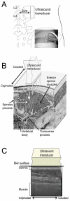
Methods: In each of 107 human subjects (60 with LBP and 47 without LBP), parasagittal ultrasound images were acquired bilaterally centered on a point 2 cm lateral to the midpoint of the L2-3 interspinous ligament. The outcome measures based on these images were subcutaneous and perimuscular connective tissue thickness and echogenicity measured by ultrasound.
Results: There were no significant differences in age, sex, body mass index (BMI) or activity levels between LBP and No-LBP groups. Perimuscular thickness and echogenicity were not correlated with age but were positively correlated with BMI. The LBP group had approximately 25% greater perimuscular thickness and echogenicity compared with the No-LBP group (ANCOVA adjusted for BMI, p<0.01 and p<0.001 respectively).
Conclusion: This is the first report of abnormal connective tissue structure in the lumbar region in a group of subjects with chronic or recurrent LBP. This finding was not attributable to differences in age, sex, BMI or activity level between groups. Possible causes include genetic factors, abnormal movement patterns and chronic inflammation.
Figures


Similar articles
-
Reduced thoracolumbar fascia shear strain in human chronic low back pain.BMC Musculoskelet Disord. 2011 Sep 19;12:203. doi: 10.1186/1471-2474-12-203. BMC Musculoskelet Disord. 2011. PMID: 21929806 Free PMC article.
-
Ultrasound evidence of altered lumbar fascia in patients with low back pain.Clin Anat. 2023 Jan;36(1):36-41. doi: 10.1002/ca.23964. Epub 2022 Oct 24. Clin Anat. 2023. PMID: 36199243
-
Structural remodeling of the lumbar multifidus, thoracolumbar fascia and lateral abdominal wall perimuscular connective tissues: A search for its potential determinants.J Anat. 2021 Mar;238(3):536-550. doi: 10.1111/joa.13330. Epub 2020 Oct 18. J Anat. 2021. PMID: 33070313 Free PMC article.
-
Structural remodelling of the lumbar multifidus, thoracolumbar fascia and lateral abdominal wall perimuscular connective tissues: A cross-sectional and comparative ultrasound study.J Bodyw Mov Ther. 2020 Oct;24(4):293-302. doi: 10.1016/j.jbmt.2020.07.009. Epub 2020 Aug 4. J Bodyw Mov Ther. 2020. PMID: 33218526
-
Effect of MELT method on thoracolumbar connective tissue: The full study.J Bodyw Mov Ther. 2017 Jan;21(1):179-185. doi: 10.1016/j.jbmt.2016.05.010. Epub 2016 Jun 3. J Bodyw Mov Ther. 2017. PMID: 28167175 Clinical Trial.
Cited by
-
Combined Oxygen-Ozone and Porcine Injectable Collagen Therapies Boosting Efficacy in Low Back Pain and Disability.Diagnostics (Basel). 2024 Oct 29;14(21):2411. doi: 10.3390/diagnostics14212411. Diagnostics (Basel). 2024. PMID: 39518378 Free PMC article.
-
Effects of percussive massage therapy on fascia echo intensity and fascia thickness in firefighters with chronic non-specific low back pain: a randomized controlled trial.BMC Complement Med Ther. 2024 Nov 8;24(1):390. doi: 10.1186/s12906-024-04687-9. BMC Complement Med Ther. 2024. PMID: 39516833 Free PMC article. Clinical Trial.
-
Ultrasound-Guided Fascial Hydrodissection with Eperisone: A Retrospective Study on Efficacy and Safety in Lumbodorsal Fasciitis Treatment.Med Sci Monit. 2024 Nov 1;30:e945874. doi: 10.12659/MSM.945874. Med Sci Monit. 2024. PMID: 39482828 Free PMC article.
-
Ultrasound elastography of back muscle biomechanical properties: a systematic review and meta-analysis of current methods.Insights Imaging. 2024 Aug 14;15(1):206. doi: 10.1186/s13244-024-01785-7. Insights Imaging. 2024. PMID: 39143409 Free PMC article. Review.
-
Changes of trunk muscle stiffness in individuals with low back pain: a systematic review with meta-analysis.BMC Musculoskelet Disord. 2024 Feb 19;25(1):155. doi: 10.1186/s12891-024-07241-3. BMC Musculoskelet Disord. 2024. PMID: 38373986 Free PMC article.
References
-
- De Luca CJ. Low back pain: a major problem with low priority. J Rehabil Res Dev. 1997;34(4):vii–viii. - PubMed
-
- Van Nieuwenhuyse A, Fatkhutdinova L, Verbeke G, Pirenne D, Johannik K, Somville PR, Mairiaux P, Moens GF, Masschelein R. Risk factors for first-ever low back pain among workers in their first employment 10.1093/occmed/kqh091. Occup Med (Lond) 2004;54(8):513–519. doi: 10.1093/occmed/kqh091. - DOI - PubMed
Publication types
MeSH terms
Grants and funding
LinkOut - more resources
Full Text Sources
Other Literature Sources
Miscellaneous


