iBet uBet web content aggregator. Adding the entire web to your favor.


Link to original content: http://www.ncbi.nlm.nih.gov/pubmed/19648118
Specification of neuronal polarity regulated by local translation of CRMP2 and Tau via the mTOR-p70S6K pathway - PubMed Skip to main page content
U.S. flag

An official website of the United States government

Dot gov

The .gov means it’s official.
Federal government websites often end in .gov or .mil. Before sharing sensitive information, make sure you’re on a federal government site.

Https

The site is secure.
The https:// ensures that you are connecting to the official website and that any information you provide is encrypted and transmitted securely.

Access keys NCBI Homepage MyNCBI Homepage Main Content Main Navigation
. 2009 Oct 2;284(40):27734-45.
doi: 10.1074/jbc.M109.008177. Epub 2009 Jul 31.

Specification of neuronal polarity regulated by local translation of CRMP2 and Tau via the mTOR-p70S6K pathway

Affiliations

Specification of neuronal polarity regulated by local translation of CRMP2 and Tau via the mTOR-p70S6K pathway

Tsuyoshi Morita et al. J Biol Chem. .

Abstract

Mammalian target of rapamycin (mTOR) is an important regulator of neuronal development and functions. Although it was reported recently that mTOR signaling is critical for neuronal polarity, the underlying mechanism remains unclear. Here, we describe the molecular pathway of mTOR-dependent axon specification, in which the collapsing response mediator protein 2 (CRMP2) and Tau are major downstream targets. The activity of mTOR effector 70-kDa ribosomal protein S6 kinase (p70S6K) specifically increases in the axon during neuronal polarity formation. The mTOR inhibitor rapamycin suppresses the translation of some neuronal polarity proteins, including CRMP2 and Tau, thereby inhibiting axon formation. In contrast, constitutively active p70S6K up-regulates the translation of these molecules, thus inducing multiple axons. Exogenous CRMP2 and Tau facilitate axon formation, even in the presence of rapamycin. In the 5'-untranslated region of Tau and CRMP2 mRNAs, we identified a 5'-terminal oligopyrimidine tract, which mediates mTOR-governed protein synthesis. The 5'-terminal oligopyrimidine tract sequences of CRMP2 and Tau mRNAs strongly contribute to the up-regulation of their translation in the axon in response to the axonal activation of the mTOR-p70S6K pathway. Taken together, we conclude that the local translation of CRMP2 and Tau, regulated by mTOR-p70S6K, is critical for the specification of neuronal polarity.


Figures
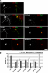
FIGURE 1.
FIGURE 1.
The mTOR pathway regulates neuronal polarity. A, isolated hippocampal neurons were transfected with pCAGGS-EGFP, pCAGGS-FLAG-CA-Rheb, pCAGGS-FLAG-CA-S6K, pCAGGS-FLAG-eIF-4E, pCAGGS-FLAG-DN-Rheb, pCAGGS-FLAG-DN-S6K, or pCAGGS-FLAG-4EBP1. After 3 DIV, the neurons were fixed and stained with anti-GFP (white), anti-FLAG (white), anti-MAP2 (dendrite marker, red), and anti-Tau1 (axon marker, green) antibodies. Bar, 50 μm. B, from the immunostained images shown in A, the transfected neurons were classified according to their polarity: no axon (white bars), single axon (black bars), or multiple axons (gray bars). Statistical analysis was carried out for four independent experiments.
FIGURE 2.
FIGURE 2.
p70S6K is critical for mTOR-regulated neuronal polarity. A, isolated hippocampal neurons were transfected with pCAGGS-EGFP, pCAGGS-FLAG-CA-Rheb, or pCAGGS-FLAG-CA-S6K and incubated with or without 100 nm rapamycin (rapa). After 3 DIV, the neurons were fixed and stained with anti-GFP (white), anti-FLAG (white), anti-MAP2 (red), and anti-Tau1 (green) antibodies. Bar, 50 μm. B, from the immunostained images shown in A, the transfected neurons were classified according to their polarity: no axon (white bars), single axon (black bars), or multiple axons (gray bars). Statistical analysis was carried out for four independent experiments. C, hippocampal neurons at 3 DIV were fixed and stained with anti-phosphorylated p70S6K (Thr421/Ser424) (p-S6K), anti-p70S6K (S6K), anti-phosphorylated 4E-BP1 (Ser65) (p-4E-BP1), and anti-4E-BP1 (4E-BP1) antibodies. The fluorescent intensities of immunostained images of the dendrites (red) and axons (green) were measured, respectively, and are presented in the graph. Bar, 50 μm.
FIGURE 3.
FIGURE 3.
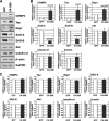
Translational regulation of neuronal polarity proteins by the mTOR-S6K pathway. A, hippocampal neurons were transfected with pCAGGS-EGFP or pCAGGS-FLAG-CA-S6K by nucleofection and cultured for 2 days. The total proteins were then extracted from the neurons. Neuronal polarity proteins were detected by Western blot analyses using the indicated antibodies. B, the amounts of proteins were determined by densitometry and statistically analyzed by Student's t test. The quantification of each protein was normalized to GAPDH. Statistical analyses were carried out for three independent experiments. C, total RNAs were extracted from the GFP- or CA-S6K-transfected neurons, and expression of mRNAs for neuronal polarity genes was detected by real time RT-PCR using gene-specific PCR primers. Statistical analyses were carried out for three independent experiments.
FIGURE 4.
FIGURE 4.
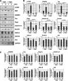
Effect of rapamycin on the translation of neuronal polarity proteins. A, hippocampal neurons were cultured with or without 100 nm rapamycin (rapa) for 1–3 days. The total proteins were then extracted from the neurons. Neuronal polarity proteins were detected by Western blot analyses using the indicated antibodies. B, the amounts of proteins were determined by densitometry and statistically analyzed by Student's t test. The quantification of phosphorylated p70S6K was normalized to total p70S6K. The quantification of other protein was normalized to GAPDH. Statistical analyses were carried out for three independent experiments. C, total RNAs were extracted from the rapamycin-treated neurons, and expression of mRNAs for neuronal polarity genes was detected by real time RT-PCR using gene-specific PCR primers. Statistical analyses were carried out for three independent experiments.
FIGURE 5.
FIGURE 5.
CA-S6K rescues the rapamycin-induced reduction in the expression of polarity proteins. A, hippocampal neurons were transfected with pCAGGS-EGFP or pCAGGS-FLAG-CA-S6K by nucleofection and cultured for 2.5 days with or without 100 nm rapamycin (rapa). The total proteins were then extracted from the neurons. CRMP2, Tau, and Rap1 proteins were detected by Western blot analyses. B, the amounts of proteins were determined by densitometry and statistically analyzed by Student's t test. The quantification of each protein was normalized to GAPDH. Statistical analyses were carried out for three independent experiments.
FIGURE 6.
FIGURE 6.
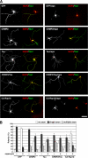
Overexpression of CRMP2 and Tau rescues the rapamycin-induced polarity defect. A, hippocampal neurons were transfected with pCAGGS-EGFP, pCAGGS-FLAG-CRMP2, pCAGGS-FLAG-Tau, and/or pCAGGS-FLAG-CA-Rap1b and cultured with or without 100 nm rapamycin (rapa) for 3 days. The cells were fixed and stained with anti-GFP (white), anti-FLAG (white), anti-MAP2 (red), and anti-Tau1 (green) antibodies. Bar, 50 μm. B, from the immunostained images shown in A, the transfected neurons were classified according to their polarity: no axon (white bars), single axon (black bars), or multiple axons (gray bars). Statistical analysis was carried out for four independent experiments.
FIGURE 7.
FIGURE 7.
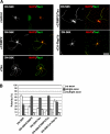
Overexpression of CRMP2 and Tau rescues the DN-S6K-induced polarity defect. A, hippocampal neurons were transfected with pCAGGS-HA-DN-S6K, pCAGGS-FLAG-CRMP2, pCAGGS-FLAG-Tau, and/or pCAGGS-FLAG-CA-Rap1b and cultured for 3 days. The cells were fixed and stained with anti-HA (white), anti-MAP2 (red), and anti-Tau1 (green) antibodies. Bar, 50 μm. B, from the immunostained images shown in A, the transfected neurons were classified according to their polarity: no axon (white bars), single axon (black bars), or multiple axons (gray bars). Statistical analysis was carried out for four independent experiments.
FIGURE 8.
FIGURE 8.
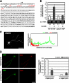
Axonal translation of CRMP2 was regulated by the mTOR-p70S6K pathway through its 5′-TOP tract. A, sequences of the 5′-UTR and promoter region of the CRMP2 gene. Transcriptional start sites of the CRMP2 mRNA (black and red arrows) were determined by RLM-5′-RACE. Red arrows indicate the transcriptional start sites conforming to the rule for 5′-TOP mRNAs. Underlined letters indicate the pyrimidine-rich region. Bold letters indicate the translational start codon (ATG). B, a luciferase (luc) reporter assay performed using pGL4.14-CRMP2 or pGL4.14-CRMP2 (Δ5′-TOP) vectors. pCAGGS-FLAG-CA-S6K was co-transferred to activate the 5′-TOP-mediated translation. Total RNAs were extracted from the transfected cells, and expression levels of ectopic luciferase mRNA were determined by RT-PCR. The numbers indicate the relative luciferase expression levels, which were quantitated by densitometry and normalized to β-galactosidase mRNA. C, localization of endogenous CRMP2 protein. Hippocampal neurons at 3 DIV were fixed and stained with anti-CRMP2 antibody. The fluorescence intensities of the immunostained images of dendrites (red) and axons (green) were measured, respectively, and are presented in the graph. Bar, 50 μm. D, hippocampal neurons co-transfected with pCAGGS-EGFP and pGL4.22-CRMP2-myr-Luc and cultured with or without 100 nm rapamycin. After 3 DIV, the neurons were fixed and stained with anti-GFP (green) and anti-luciferase (red) antibodies. The fluorescence intensities of the immunostained images of the dendrites and axon were measured, respectively, and are presented in the graph. Bar, 50 μm.
FIGURE 9.
FIGURE 9.
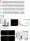
Axonal translation of Tau was also regulated by the mTOR-p70S6K pathway through its 5′-TOP sequence. A, alignment of 5′-UTR sequences of the mouse, rat, and human Tau mRNAs. Red box indicates the 5′-TOP sequence. Blue box indicates translational start codon (ATG). Asterisks indicate residues conserved in all three species. B, a luciferase (luc) reporter assay performed using pGL4.14-Tau or pGL4.14-Tau (mut 5′-TOP) vectors. pCAGGS-FLAG-CA-S6K was co-transferred to activate the 5′-TOP-mediated translation. Total RNAs were extracted from the transfected cells, and expression levels of ectopic luciferase mRNA were determined by RT-PCR. The numbers indicate the relative luciferase expression levels, which were quantitated by densitometry and normalized to β-galactosidase mRNA. C, localization of endogenous Tau protein. Hippocampal neurons at 3 DIV were fixed and stained with anti-Tau antibody. The fluorescence intensities of the immunostained images of dendrites (red) and axons (green) were measured, respectively, and are presented in the graph. Bar, 50 μm. D, hippocampal neurons were co-transfected with pCAGGS-EGFP and pGL4.22-Tau-myr-Luc and cultured with or without 100 nm rapamycin. After 3 DIV, the neurons were fixed and stained with anti-GFP (green) and anti-luciferase (red) antibodies. The fluorescence intensities of the immunostained images of the dendrites and axon were measured, respectively, and are presented in the graph. Bar, 50 μm.

Similar articles

Cited by

References

    1. Dotti C. G., Sullivan C. A., Banker G. A. (1988) J. Neurosci. 8, 1454–1468 - PMC - PubMed
    1. Craig A. M., Banker G. (1994) Annu. Rev. Neurosci. 17, 267–310 - PubMed
    1. Arimura N., Kaibuchi K. (2007) Nat. Rev. Neurosci. 8, 194–205 - PubMed
    1. Hay N., Sonenberg N. (2004) Genes Dev. 18, 1926–1945 - PubMed
    1. Swiech L., Perycz M., Malik A., Jaworski J. (2008) Biochim. Biophys. Acta 1784, 116–132 - PubMed

Publication types

MeSH terms