Does breast cancer start in the womb?
- PMID: 18226065
- PMCID: PMC2817934
- DOI: 10.1111/j.1742-7843.2007.00165.x
Does breast cancer start in the womb?
Abstract
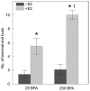
Perturbations in the foetal environment predispose individuals to diseases that become apparent in adulthood. These findings prompted researchers to hypothesize that foetal exposure to environmental oestrogens may play a role in the increased incidence of breast cancer observed in European and US populations over the last 50 years. There is widespread human exposure to bisphenol A, an oestrogenic compound that leaches from dental materials and consumer products. In CD-1 mice, perinatal exposure to environmentally relevant bisphenol A levels induced alterations of the mammary gland architecture. Bisphenol A increased the number of terminal end buds at puberty and terminal ends at 6 months of age and increased ductal lateral branching at 4 months of age. Exposed mice also showed an enhanced sensitivity to oestradiol when ovariectomized prior to puberty. All these parameters are associated in human beings with an increased risk for developing breast cancer. To assess whether bisphenol A induces mammary gland neoplasia, we chose a rat model because it more closely mimics the human disease than mouse models. Examination of the mammary glands of Wistar/Furth rats during early adulthood revealed that gestational exposure to bisphenol A induced the development of pre-neoplastic lesions and carcinoma in situ in the absence of any additional treatment aimed at increasing tumour development. Emerging epidemiological data reveal an increased incidence of breast cancer in women exposed to diethylstilboestrol during gestation. Hence, both animal experiments and epidemiological data strengthen the hypothesis that foetal exposure to xenooestrogens may be an underlying cause of the increased incidence of breast cancer observed over the last 50 years.
Figures


Similar articles
-
Perinatal exposure to bisphenol-A alters peripubertal mammary gland development in mice.Endocrinology. 2005 Sep;146(9):4138-47. doi: 10.1210/en.2005-0340. Epub 2005 May 26. Endocrinology. 2005. PMID: 15919749 Free PMC article.
-
Induction of mammary gland ductal hyperplasias and carcinoma in situ following fetal bisphenol A exposure.Reprod Toxicol. 2007 Apr-May;23(3):383-90. doi: 10.1016/j.reprotox.2006.10.002. Epub 2006 Oct 24. Reprod Toxicol. 2007. PMID: 17123778 Free PMC article.
-
Prenatal bisphenol A exposure induces preneoplastic lesions in the mammary gland in Wistar rats.Environ Health Perspect. 2007 Jan;115(1):80-6. doi: 10.1289/ehp.9282. Environ Health Perspect. 2007. PMID: 17366824 Free PMC article.
-
Estrogens in the wrong place at the wrong time: Fetal BPA exposure and mammary cancer.Reprod Toxicol. 2015 Jul;54:58-65. doi: 10.1016/j.reprotox.2014.09.012. Epub 2014 Sep 30. Reprod Toxicol. 2015. PMID: 25277313 Free PMC article. Review.
-
Endocrine-disrupting compounds and mammary gland development: early exposure and later life consequences.Endocrinology. 2006 Jun;147(6 Suppl):S18-24. doi: 10.1210/en.2005-1131. Epub 2006 May 11. Endocrinology. 2006. PMID: 16690811 Review.
Cited by
-
Biological Basis of Breast Cancer-Related Disparities in Precision Oncology Era.Int J Mol Sci. 2024 Apr 8;25(7):4113. doi: 10.3390/ijms25074113. Int J Mol Sci. 2024. PMID: 38612922 Free PMC article. Review.
-
Breast Cancer Exposomics.Life (Basel). 2024 Mar 18;14(3):402. doi: 10.3390/life14030402. Life (Basel). 2024. PMID: 38541726 Free PMC article. Review.
-
Integrated Genomic and Bioinformatics Approaches to Identify Molecular Links between Endocrine Disruptors and Adverse Outcomes.Int J Environ Res Public Health. 2022 Jan 5;19(1):574. doi: 10.3390/ijerph19010574. Int J Environ Res Public Health. 2022. PMID: 35010832 Free PMC article. Review.
-
Implication of the Strand-Specific Imprinting and Segregation Model: Integrating in utero Hormone Exposure, Stem Cell and Lateral Asymmetry Hypotheses in Breast Cancer Aetiology.Hereditary Genet. 2013;2013(Suppl 2):005. doi: 10.4172/2161-1041.s2-005. Epub 2013 Aug 13. Hereditary Genet. 2013. PMID: 34589269 Free PMC article.
-
Bisphenol-A exposure and risk of breast and prostate cancer in the Spanish European Prospective Investigation into Cancer and Nutrition study.Environ Health. 2021 Aug 16;20(1):88. doi: 10.1186/s12940-021-00779-y. Environ Health. 2021. PMID: 34399780 Free PMC article.
References
-
- Hahn WC, Weinberg RA. Modelling the molecular circuitry of cancer. Nat Rev Cancer. 2002;2:331–42. - PubMed
-
- Sonnenschein C, Soto AM. The Society of Cells: Cancer and Control of Cell Proliferation. Springer Verlag; New York: 1999.
-
- Maffini MV, Soto AM, Calabro JM, Ucci AA, Sonnenschein C. The stroma as a crucial target in rat mammary gland carcinogenesis. J Cell Sci. 2004;117:1495–502. - PubMed
-
- Gilbert SF. Mechanisms for the environmental regulation of gene expression: ecological aspects of animal development. J Biosci. 2005;30:65–74. - PubMed
-
- Colborn T, Clement C, editors. Chemically Induced Alterations in Sexual and Functional Development: The Wildlife/Human Connection. Princeton Scientific Publishing; Princeton, NJ: 1992.
Publication types
MeSH terms
Substances
Grants and funding
LinkOut - more resources
Full Text Sources


