The therapeutic role of 5-HT1A and 5-HT2A receptors in depression
- PMID: 15309042
- PMCID: PMC446220
The therapeutic role of 5-HT1A and 5-HT2A receptors in depression
Abstract
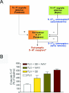
The selective serotonin reuptake inhibitors (SSRIs) are the most frequently prescribed antidepressant drugs, because they are well tolerated and have no severe side effects. They rapidly block serotonin (5-HT) reuptake, yet the onset of their therapeutic action requires weeks of treatment. This delay is the result of presynaptic and postsynaptic adaptive mechanisms secondary to reuptake inhibition. The prevention of a negative feedback mechanism operating at the 5-HT autoreceptor level enhances the neurochemical and clinical effects of SSRIs. The blockade of 5-HT2A receptors also seems to improve the clinical effects of SSRIs. These receptors are located postsynaptically to 5-HT axons, mainly in the neocortex. Pyramidal neurons in the prefrontal cortex are particularly enriched in 5-HT2A receptors. Their blockade may affect the function of prefrontal-subcortical circuits, an effect that probably underlies the beneficial effects of the addition of atypical antipsychotic drugs, which are 5-HT2A receptor antagonists, to SSRIs in treatment-resistant patients.
Les inhibiteurs spécifiques du recaptage de la sérotonine (ISRS) sont le plus souvent prescrits comme antidépresseurs parce qu'ils sont bien tolérés et ne produisent pas d'effets secondaires graves. Ils bloquent rapidement le recaptage de la sérotonine (5-HT), mais il faut des semaines de traitement pour que leur effet thérapeutique se fasse sentir. Ce retard est attribuable à des mécanismes d'adaptation présynaptiques et postsynaptiques secondaires à l'inhibition du recaptage. La prévention d'un mécanisme de rétroaction négative fonctionnant au niveau des autorécepteurs de la 5-HT améliore les effets neurochimiques et cliniques des ISRS. Le blocage des récepteurs de la 5-HT2A semble aussi améliorer l'effet clinique des ISRS. Ces récepteurs sont situés dans la région postsynatique par rapport aux axones de 5-HT, principalement dans le néocortex. Les neurones pyramidaux du cortex préfrontal sont particulièrement riches en récepteurs de la 5-HT2A. Leur blocage peut avoir, sur le fonctionnement des circuits préfrontaux-sous-corticaux, un effet qui sous-tend probablement les effets bénéfiques de l'ajout d'antipsychotiques atypiques, qui sont des antagonistes des récepteurs de la 5-HT2A, chez les patients résistants au traitement aux ISRS.
Figures


Similar articles
-
Other Antidepressants.Handb Exp Pharmacol. 2019;250:325-355. doi: 10.1007/164_2018_167. Handb Exp Pharmacol. 2019. PMID: 30194544
-
[Interest of the use of pindolol in the treatment of depression: review].Encephale. 2003 Jul-Aug;29(4 Pt 1):338-50. Encephale. 2003. PMID: 14615704 Review. French.
-
Acute effects of combining citalopram and pindolol on regional brain serotonin synthesis in sham operated and olfactory bulbectomized rats.Neurochem Int. 2009 Mar-Apr;54(3-4):161-71. doi: 10.1016/j.neuint.2008.08.012. Epub 2008 Nov 27. Neurochem Int. 2009. PMID: 19073228 Free PMC article.
-
Blockade of 5-HT1A receptors by (+/-)-pindolol potentiates cortical 5-HT outflow, but not antidepressant-like activity of paroxetine: microdialysis and behavioral approaches in 5-HT1A receptor knockout mice.Neuropsychopharmacology. 2006 Oct;31(10):2162-72. doi: 10.1038/sj.npp.1301019. Epub 2006 Jan 25. Neuropsychopharmacology. 2006. PMID: 16452992
-
Pharmacology of rapid-onset antidepressant treatment strategies.J Clin Psychiatry. 2001;62 Suppl 15:12-7. J Clin Psychiatry. 2001. PMID: 11444761 Review.
Cited by
-
Linking the microarchitecture of neurotransmitter systems to large-scale MEG resting state networks.iScience. 2024 Oct 9;27(11):111111. doi: 10.1016/j.isci.2024.111111. eCollection 2024 Nov 15. iScience. 2024. PMID: 39524335 Free PMC article.
-
Gepirone: A New Extended-Release Oral Selective Serotonin Receptor Agonist for Major Depressive Disorder.J Pharm Technol. 2024 Oct;40(5):230-235. doi: 10.1177/87551225241269179. Epub 2024 Aug 13. J Pharm Technol. 2024. PMID: 39493804 Review.
-
Recent advances in the synthesis of antidepressant derivatives: pharmacologic insights for mood disorders.3 Biotech. 2024 Nov;14(11):260. doi: 10.1007/s13205-024-04104-5. Epub 2024 Oct 5. 3 Biotech. 2024. PMID: 39376479 Review.
-
Discovery of ITI-333, a Novel Orally Bioavailable Molecule Targeting Multiple Receptors for the Treatment of Pain and Other Disorders.J Med Chem. 2024 Jun 13;67(11):9355-9373. doi: 10.1021/acs.jmedchem.4c00480. Epub 2024 May 28. J Med Chem. 2024. PMID: 38805667 Free PMC article.
-
Pharmacologic profile of ITI-333: a novel molecule for treatment of substance use disorders.Psychopharmacology (Berl). 2024 Jul;241(7):1477-1490. doi: 10.1007/s00213-024-06578-w. Epub 2024 May 6. Psychopharmacology (Berl). 2024. PMID: 38710856 Free PMC article.
References
-
- Murray CGL, Lopez AD. Alternative projections of mortality and disability by cause 1990-2020: Global Burden of Disease Study. Lancet 1997;349:1498-504. - PubMed
-
- Danish University Antidepressant Group. Citalopram: clinical effect profile in comparison with clomipramine. A controlled multicenter study. Psychopharmacology 1986;90:131-8. - PubMed
-
- Danish University Antidepressant Group. Paroxetine: a selective serotonin reuptake inhibitor showing better tolerance, but weaker antidepressant effect than clomipramine in a controlled multicenter study. J Affect Disord 1990;18:289-99. - PubMed
-
- Blier P, Ward NM. Is there a role for 5-HT1A agonists in the treatment of depression? Biol Psychiatry 2003;53:93-103. - PubMed
Publication types
MeSH terms
Substances
LinkOut - more resources
Full Text Sources
Other Literature Sources

