iBet uBet web content aggregator. Adding the entire web to your favor.


Link to original content: http://pubmed.ncbi.nlm.nih.gov/39519499/
In Vitro Assessment of the Neuroprotective Effects of Pomegranate (Punica granatum L.) Polyphenols Against Tau Phosphorylation, Neuroinflammation, and Oxidative Stress - PubMed Skip to main page content
U.S. flag

An official website of the United States government

Dot gov

The .gov means it’s official.
Federal government websites often end in .gov or .mil. Before sharing sensitive information, make sure you’re on a federal government site.

Https

The site is secure.
The https:// ensures that you are connecting to the official website and that any information you provide is encrypted and transmitted securely.

Access keys NCBI Homepage MyNCBI Homepage Main Content Main Navigation
. 2024 Oct 28;16(21):3667.
doi: 10.3390/nu16213667.

In Vitro Assessment of the Neuroprotective Effects of Pomegranate (Punica granatum L.) Polyphenols Against Tau Phosphorylation, Neuroinflammation, and Oxidative Stress

Affiliations

In Vitro Assessment of the Neuroprotective Effects of Pomegranate (Punica granatum L.) Polyphenols Against Tau Phosphorylation, Neuroinflammation, and Oxidative Stress

Mehdi Alami et al. Nutrients. .

Abstract

Background: Oxidative stress and chronic inflammation, at both the systemic and the central level, are critical early events in atherosclerosis and Alzheimer's disease (AD).

Purpose: To investigate the oxidative stress-, inflammation-, and Tau-phosphorylation-lowering effects of pomegranate polyphenols (PPs) (punicalagin, ellagic acid, peel, and aril extracts).

Methods: We used flow cytometry to quantify the protein expression of proinflammatory cytokines (IL-1β) and anti-inflammatory mediators (IL-10) in THP-1 macrophages, as well as M1/M2 cell-specific marker (CD86 and CD163) expression in human microglia HMC3 cells. The IL-10 protein expression was also quantified in U373-MG human astrocytes. The effect of PPs on human amyloid beta 1-42 (Aβ1-42)-induced oxidative stress was assessed in the microglia by measuring ROS generation and lipid peroxidation, using 2',7'-dichlorofluorescein diacetate (DCFH-DA) and thiobarbituric acid reactive substance (TBARS) tests, respectively. Neuronal viability and cell apoptotic response to Aβ1-42 toxicity were assayed using the MTT (3-(4,5-dimethylthiazolyl-2)-2,5-diphenyltetrazolium bromide) assay and the annexin-V-FITC apoptosis detection kit, respectively. Finally, flow cytometry analysis was also performed to evaluate the ability of PPs to modulate Aβ1-42-induced Tau-181 phosphorylation (pTau-181).

Results: Our data indicate that PPs are significantly (p < 0.05) effective in countering Aβ1-42-induced inflammation through increasing the anti-inflammatory cytokines (IL-10) in U373-MG astrocytes and THP1 macrophages and decreasing proinflammatory marker (IL-1β) expression in THP1 macrophages. The PPs were also significantly (p < 0.05) effective in inducing the phenotypic transition of THP-1 macrophages and microglial cells from M1 to M2 by decreasing CD86 and increasing CD163 surface receptor expression. Moreover, our treatments have a significant (p < 0.05) beneficial impact on oxidative stress, illustrated in the reduction in TBARS and ROS generation. Our treatments have significant (p < 0.05) cell viability improvement capacities and anti-apoptotic effects on human H4 neurons. Furthermore, our results suggest that Aβ1-42 significantly (p < 0.05) increases pTau-181. This effect is significantly (p < 0.05) attenuated by arils, peels, and punicalagin and drastically reduced by the ellagic acid treatment.

Conclusion: Overall, our results attribute to PPs anti-inflammatory, antioxidant, anti-apoptotic, and anti-Tau-pathology potential. Future studies should aim to extend our knowledge of the potential role of PPs in Aβ1-42-induced neurodegeneration, particularly concerning its association with the tauopathy involved in AD.

Keywords: Alzheimer’s disease; amyloid-beta; ellagic acid; microglia; neuroinflammation; oxidative stress; phospho-Tau-181; pomegranate (Punica granatum L.); punicalagin.


Conflict of interest statement

Author Ton Bunt was employed by the company Izumi Biosciences, Inc. The remaining authors declare that the research was conducted in the absence of any commercial or financial relationships that could be construed as a potential conflict of interest.

Figures

Figure 1
Figure 1
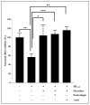
The modulatory effects of peels, arils, punicalagin, and ellagic acid on human Aβ1-42-induced neuronal cytotoxicity. Neuronal H4 cells were exposed, or not, to 20 µg/mL of Aβ1-42 in the presence or absence of the above treatments for 24 h. (A) represents the effect of Aβ1-42 on the neuronal cells viability. (B) illustrates the effects of PPs (peels: 200 µg/mL; arils: 200 µg/mL; punicalagin: 50 µg/mL; and ellagic acid: 50 µM) on the Aβ1-42-related cytotoxicity. Data are expressed as means ± SEM. (*) vs. Aβ1-42: * p < 0.05, ** p < 0.01.
Figure 2
Figure 2
Elacridar improves the effect of pomegranate-arils-rich phenolic extract and punicalagin polyphenols against human-Aβ1-42-induced neuronal death. H4 cells were stimulated, or not, with Aβ1-42 (20 µg/mL) for 24 h, and simultaneously treated with pomegranate arils extract (200 µg/mL) or punicalagin (50 µg/mL) in the presence of elacridar (500 ng/mL). Data are expressed as means ± SEM. (*) vs. Aβ1-42: * p < 0.05, ** p < 0.01, *** p < 0.001.
Figure 3
Figure 3
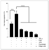
The neuronal-survival-enhancement effect of pomegranate peels (200 µg/mL), arils (200 µg/mL), punicalagin (50 µg/mL), and ellagic acid (50 µM). The H4 neurons were incubated with or without 15 µg/mL of human Aβ1-42 for 24 h, in the presence or absence of the above treatments. The obtained results are expressed as mean ± SEM. (*) vs. Aβ1-42: *** p < 0.001; **** p < 0.0001.
Figure 4
Figure 4
Pomegranate polyphenols reduce ROS generation in human microglia HMC3 cells. Cells were pretreated with human Aβ1-42 (5 µg/mL) in the presence, or not, of different concentrations of pomegranate peels (A), arils (B), punicalagin (C) and ellagic acid (D). The obtained data are presented as mean ± SEM. (*) vs. Aβ1-42: * p < 0.05.
Figure 5
Figure 5
The protective effect of pomegranate polyphenols on lipid peroxidation. HMC3 microglia cells were pretreated (24 h) with peel (A) and aril (B) extracts, punicalagin (C), and ellagic acid (D) compounds before their stimulation with TBHP (200 µM) for 1 h. The data are presented as mean ± SEM of at least three independent experiments. * p < 0.05; ** p < 0.01; *** p < 0.001.
Figure 6
Figure 6
The modulatory effects of peels, arils, punicalagin, and ellagic acid on M1/M2 polarization of HMC3 microglia cells. HMC3 cells were stimulated by 1 µg/mL of LPS and cotreated simultaneously with pomegranate peels, arils, punicalagin, and ellagic acid for 24 h. (a) represents the effect of PPs on CD86 expression. (14) illustrates respectively, the effect of pomegranate peels, arils, punicalagin and ellagic acid on CD86 expression. (b) illustrates the impact of PPs on CD163 receptor expression. (14) represents the effect of pomegranate peels, arils, punicalagin, and ellagic acid on gene expression of CD163. (*) vs. LPS: * p< 0.05; ** p< 0.01; *** p< 0.001; **** p< 0.0001.
Figure 6
Figure 6
The modulatory effects of peels, arils, punicalagin, and ellagic acid on M1/M2 polarization of HMC3 microglia cells. HMC3 cells were stimulated by 1 µg/mL of LPS and cotreated simultaneously with pomegranate peels, arils, punicalagin, and ellagic acid for 24 h. (a) represents the effect of PPs on CD86 expression. (14) illustrates respectively, the effect of pomegranate peels, arils, punicalagin and ellagic acid on CD86 expression. (b) illustrates the impact of PPs on CD163 receptor expression. (14) represents the effect of pomegranate peels, arils, punicalagin, and ellagic acid on gene expression of CD163. (*) vs. LPS: * p< 0.05; ** p< 0.01; *** p< 0.001; **** p< 0.0001.
Figure 7
Figure 7
The effect of pomegranate polyphenols on CD86 (a) and CD163 (b) protein expression. Human-THP-1-derived macrophages were stimulated by LPS (1 µg/mL) and cotreated with pomegranate peels (1), aril-rich phenolic extracts (2), punicalagin (3), and ellagic acid (4), for 24 h. (*) vs. LPS: * p< 0.05; ** p< 0.01; *** p< 0.001.
Figure 7
Figure 7
The effect of pomegranate polyphenols on CD86 (a) and CD163 (b) protein expression. Human-THP-1-derived macrophages were stimulated by LPS (1 µg/mL) and cotreated with pomegranate peels (1), aril-rich phenolic extracts (2), punicalagin (3), and ellagic acid (4), for 24 h. (*) vs. LPS: * p< 0.05; ** p< 0.01; *** p< 0.001.
Figure 8
Figure 8
The bioeffects of pomegranate polyphenols on gene expression of IL-10. The U373-MG human astrocytes were stimulated by 1 µg/mL of LPS and cotreated simultaneously overnight with pomegranate peels (A), arils, (B) punicalagin (C), and ellagic acid (D) at different concentrations. (*) vs. LPS: ** p< 0.01; *** p< 0.001.
Figure 9
Figure 9
The modulatory effects of pomegranate peels (1), arils (2), punicalagin (3), and ellagic acid (4) on IL-10 (a) and IL-1β (b) protein expression in THP-1-derived macrophages. Cells were stimulated by 1 µg/mL of LPS and co-treated simultaneously with pomegranate polyphenols for 24 h. (*) vs. LPS: * p < 0.05; ** p < 0.01; *** p < 0.001.
Figure 9
Figure 9
The modulatory effects of pomegranate peels (1), arils (2), punicalagin (3), and ellagic acid (4) on IL-10 (a) and IL-1β (b) protein expression in THP-1-derived macrophages. Cells were stimulated by 1 µg/mL of LPS and co-treated simultaneously with pomegranate polyphenols for 24 h. (*) vs. LPS: * p < 0.05; ** p < 0.01; *** p < 0.001.
Figure 10
Figure 10
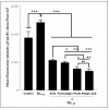
The modulatory effects of pomegranate polyphenols on human Aβ1-42-induced Tau phosphorylation at threonine 181 in H4 human neuroglioma cells. Cells were either unstimulated or stimulated with human Aβ1-42 (10 µg/mL) in the presence or absence of pomegranate polyphenols simultaneously for 24 h. Peels: 200 µg/mL; arils: 200 µg/mL; punicalagin: 50 µg/mL; ellagic acid: 50 µM. (*) vs. Aβ1-42: * p < 0.05; ** p < 0.01; *** p < 0.001.

Similar articles

References

    1. Rogers J., Luber-Narod J., Styren S.D., Civin W.H. Expression of Immune System-Associated Antigens by Cells of the Human Central Nervous System: Relationship to the Pathology of Alzheimer’s Disease. Neurobiol. Aging. 1988;9:339–349. doi: 10.1016/S0197-4580(88)80079-4. - DOI - PubMed
    1. Fiacco T.A., McCarthy K.D. Astrocyte Calcium Elevations: Properties, Propagation, and Effects on Brain Signaling. Glia. 2006;54:676–690. doi: 10.1002/glia.20396. - DOI - PubMed
    1. Floyd R.A., Hensley K. Nitrone Inhibition of Age-Associated Oxidative Damage. Ann. N. Y. Acad. Sci. 2000;899:222–237. doi: 10.1111/j.1749-6632.2000.tb06189.x. - DOI - PubMed
    1. Ventura R., Harris K.M. Three-Dimensional Relationships between Hippocampal Synapses and Astrocytes. J. Neurosci. 1999;19:6897–6906. doi: 10.1523/JNEUROSCI.19-16-06897.1999. - DOI - PMC - PubMed
    1. Boyarsky G., Ransom B., Schlue W.-R., Davis M.B.E., Boron W.F. Intracellular PH Regulation in Single Cultured Astrocytes from Rat Forebrain. Glia. 1993;8:241–248. doi: 10.1002/glia.440080404. - DOI - PubMed

MeSH terms

LinkOut - more resources