iBet uBet web content aggregator. Adding the entire web to your favor.


Link to original content: http://pubmed.ncbi.nlm.nih.gov/39167653/
Essential and multifunctional mpox virus E5 helicase-primase in double and single hexamer - PubMed Skip to main page content
U.S. flag

An official website of the United States government

Dot gov

The .gov means it’s official.
Federal government websites often end in .gov or .mil. Before sharing sensitive information, make sure you’re on a federal government site.

Https

The site is secure.
The https:// ensures that you are connecting to the official website and that any information you provide is encrypted and transmitted securely.

Access keys NCBI Homepage MyNCBI Homepage Main Content Main Navigation
. 2024 Aug 23;10(34):eadl1150.
doi: 10.1126/sciadv.adl1150. Epub 2024 Aug 21.

Essential and multifunctional mpox virus E5 helicase-primase in double and single hexamer

Affiliations

Essential and multifunctional mpox virus E5 helicase-primase in double and single hexamer

Yunxia Xu et al. Sci Adv. .

Abstract

An outbreak of mpox virus in May 2022 has spread over 110 nonpandemic regions in the world, posing a great threat to global health. Mpox virus E5, a helicase-primase, plays an essential role in DNA replication, but the molecular mechanisms are elusive. Here, we report seven structures of mpox virus E5 in a double hexamer (DH) and six in single hexamer in different conformations, indicating a rotation mechanism for helicase and a coupling action for primase. The DH is formed through the interface of zinc-binding domains, and the central channel density indicates potential double-stranded DNA (dsDNA), which helps to identify dsDNA binding residues Arg249, Lys286, Lys315, and Lys317. Our work is important not only for understanding poxviral DNA replication but also for the development of novel therapeutics for serious poxviral infections including smallpox virus and mpox virus.


Figures

Fig. 1.
Fig. 1.. Biochemical characterization of mpox virus E5.
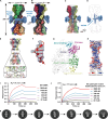
(A) Schematic diagram of domain organizations of mpox virus E5. (B) Mpox virus E5 purification by size exclusion chromatography and analysis using SDS–polyacrylamide gel electrophoresis. MW, molecular weight. (C) Mpox virus E5 binds dsDNA. The assay was performed using BLI. (D) E5 primase activity. E5 can synthesize primer RNA, when ssDNA and NTP were added. NTP, nucleoside triphosphate; dNTP, deoxyribonucleoside triphosphate.
Fig. 2.
Fig. 2.. Structure of E5 DH.
(A and B) Cryo-EM density map of E5 DH. Two hexamers are head-to-head to form DH. The primase domains are surrounded at the interface of DH. Panel (A) rotates 180° along y axis to panel (B). (C and D) Potential dsDNA density. The density is located in the channel of zinc-binding and collar domains. (E) Interaction of interface residues in the DH. (F) Density for ssDNA in the helicase domain. (G) A full-length E5 protomer structure in cartoon. (H) Electrostatic potential map of channel of DH. The channel is highly positively charged, especially in the channel of zinc-binding and collar domains. (I) Mpox virus E5 single mutant (Lys286Ala) binds dsDNA with a reduced affinity. The assay was performed using BLI. (J) Mpox virus E5 quadruple mutant (Arg249Ala/Lys286Ala/Lys315Ala/Lys317Ala) binds dsDNA with a greatly reduced affinity. The assay was performed using BLI. (K) 2D classification of DH. Different classes of DH might represent various states of DH.
Fig. 3.
Fig. 3.. Density maps of different states of E5 hexamer.
(A) Top, side, and bottom views of apo hexamer. (B) Top, side, and bottom views of ssDNA-hexamer. (C) Top, side, and bottom views of AMPPNP-ssDNA-hexamer. (D) Top, side, and bottom views of AMPPNP-ssDNA-primase-hexamer. (E) Top, side, and bottom views of ADP-ssDNA-primase-hexamer. (F) Top, side, and bottom views of 1ATP-3ADP-ssDNA-hexamer.
Fig. 4.
Fig. 4.. E5 Hexamer binds ssDNA.
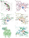
(A) Superimposition of ssDNA-hexamer and AMPPNP-ssDNA-hexamer. The ssDNA is shown in cartoon, ssDNA-hexamer in green, and AMPPNP-ssDNA-hexamer in purple. (B) ssDNA is bound by residues of AMPPNP-ssDNA-hexamer. (C) ssDNA is bound by residues of ssDNA-hexamer. (D) ssDNA is bound by residues of 1ATP/ADP-ssDNA-hexamer. (E) Human primase in complex with dsDNA (PDB code: 7JKP) is superimposed to E5 primase. The core RMSD of 2.536 Å over 166 aligned residues. (F) Potential active site of primase of E5. The residues of E5 primase are in green, while human primase is in cyan. The dATP is shown in stick and cyan.
Fig. 5.
Fig. 5.. The potential mechanism of E5.
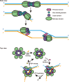
The E5 binds dsDNA at the ori to form DH. The DH uses ATP to melt the ori by grasping 3′-5′ ssDNA (C-terminal to N-terminal domains of E5). E5 separates dsDNA into ssDNA to form replication forks. E5 binds the 3′-5′ ssDNA and extrudes another ssDNA for bidirectional DNA replication. E5 uses the cycles of ATP binding, hydrolysis, and ADP release to unwind dsDNA to ssDNA.

Similar articles

References

    1. Patel S. S., Picha K. M., Structure and function of hexameric helicases. Annu. Rev. Biochem. 69, 651–697 (2000). - PubMed
    1. Yuan Z., Georgescu R., Bai L., Zhang D., Li H., O’Donnell M. E., DNA unwinding mechanism of a eukaryotic replicative CMG helicase. Nat. Commun. 11, 688 (2020). - PMC - PubMed
    1. Hamdan S. M., Richardson C. C., Motors, switches, and contacts in the replisome. Annu. Rev. Biochem. 78, 205–243 (2009). - PubMed
    1. Douglas M. E., Ali F. A., Costa A., Diffley J. F. X., The mechanism of eukaryotic CMG helicase activation. Nature 555, 265–268 (2018). - PMC - PubMed
    1. He Q., Baranovskiy A. G., Morstadt L. M., Lisova A. E., Babayeva N. D., Lusk B. L., Lim C. J., Tahirov T. H., Structures of human primosome elongation complexes. Nat. Struct. Mol. Biol. 30, 579–583 (2023). - PMC - PubMed

LinkOut - more resources