iBet uBet web content aggregator. Adding the entire web to your favor.


Link to original content: http://pubmed.ncbi.nlm.nih.gov/38793541/
Design and Application of Biosafe Coronavirus Engineering Systems without Virulence - PubMed Skip to main page content
U.S. flag

An official website of the United States government

Dot gov

The .gov means it’s official.
Federal government websites often end in .gov or .mil. Before sharing sensitive information, make sure you’re on a federal government site.

Https

The site is secure.
The https:// ensures that you are connecting to the official website and that any information you provide is encrypted and transmitted securely.

Access keys NCBI Homepage MyNCBI Homepage Main Content Main Navigation
Review
. 2024 Apr 24;16(5):659.
doi: 10.3390/v16050659.

Design and Application of Biosafe Coronavirus Engineering Systems without Virulence

Affiliations
Review

Design and Application of Biosafe Coronavirus Engineering Systems without Virulence

Guoqiang Wu et al. Viruses. .

Abstract

In the last twenty years, three deadly zoonotic coronaviruses (CoVs)-namely, severe acute respiratory syndrome coronavirus (SARS-CoV), Middle East respiratory syndrome coronavirus (MERS-CoV), and SARS-CoV-2-have emerged. They are considered highly pathogenic for humans, particularly SARS-CoV-2, which caused the 2019 CoV disease pandemic (COVID-19), endangering the lives and health of people globally and causing unpredictable economic losses. Experiments on wild-type viruses require biosafety level 3 or 4 laboratories (BSL-3 or BSL-4), which significantly hinders basic virological research. Therefore, the development of various biosafe CoV systems without virulence is urgently needed to meet the requirements of different research fields, such as antiviral and vaccine evaluation. This review aimed to comprehensively summarize the biosafety of CoV engineering systems. These systems combine virological foundations with synthetic genomics techniques, enabling the development of efficient tools for attenuated or non-virulent vaccines, the screening of antiviral drugs, and the investigation of the pathogenic mechanisms of novel microorganisms.

Keywords: application; biosafety; coronavirus; pseudovirus; trans-complementation system.


Conflict of interest statement

The authors declare no competing financial interests.

Figures

Figure 1
Figure 1
Timeline of key events related to coronavirus outbreaks. So far, seven coronaviruses that infect humans have been identified. Among them, HCoV-229E, HCoV-OC43, HCoV-NL63, and HCoV-HKU1 only cause mild respiratory diseases. Conversely, the pathogenicity and infectivity of SARS-CoV, MERS-CoV, and SARS-CoV-2 are so high that their transmission was widespread, causing deaths worldwide.
Figure 2
Figure 2
Coronavirus virion genomic structure and life cycle. (A) The structure of coronavirus particles, containing four structural proteins, namely Spike (S), Membrane (M), Nucleocapsid (N), and Envelope (E) protein, all of which are encoded within the 3′ end of the viral genome. The non-segmented positive (+)-sense single stranded RNA is surrounded by the proteins. (B) The genome structure of SARS-CoV-2, SARS-CoV, and MERS-CoV. The dotted line displays the critical difference among CoVs. (C) An overview for the life cycle of coronavirus in the host cell. When (1) recognized by cell receptors (ACE2/TMPRSS2), virus particles entered host cells by (2) direct fusion of the viral and cell host surface or through endocytosis. And (3) viral (+) ssRNA was released into the cytoplasm. (4) Non-structural proteins were translated by host ribosome with the template of genome RNA, and (5) (+) ssRNA replicated, generating the (−) ssRNA. The structural proteins were (6) translated and (7) assembled in the endoplasmic reticulum and Golgi. Finally, the virus particles were (8) matured and (9) released via exocytosis.
Figure 3
Figure 3
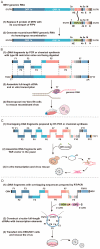
The common strategies of reverse genetics. (A) Targeted RNA recombination. The synthetic donor RNA containing the S gene of feline infectious peritonitis virus (FIPV) was transfected into mouse L2 cells, which were infected with the thermolabile MHV N gene deletion mutant. A crossover event occurs within the HE gene fragment during targeted recombination. Recombinant MHV genomic RNA containing the FIPV S gene is produced. (B) In vitro ligation. The specific sites in every fragment were recognized and cleaved by IIS restriction endonucleases, which were assembled into full-length cDNA. These were then transcribed in vitro and SARS-CoV-2 RNA was transfected in cells to rescue the virus. (C) TAR. Overlapping DNA fragments were transmitted into yeast with linearized TAR vector, and all DNA fragments were assembled by homologous recombination to generate the YAC vector containing the viral full-length cDNA. (D) CPER. Circular viral cDNA with transcription elements was constructed, which include CMV promoter, HDV ribozyme (HDVr), and transcriptional terminator sequence (polyA). The SARS-CoV-2 virus was rescued after being transfected in the package cells.
Figure 4
Figure 4
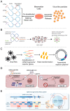
Construction strategies of biosafe coronavirus engineering systems. (A) Schematic outline of SARS-CoV-2 VLPs construction in mammalian expression system. Reproduced with permission from Ref. [80]. Copyright 2020, Frontiers Media S.A. (B) Traditional lentivirus pseudotyping method Reproduced with permission from Ref. [81]. Copyright 2023, Cell Press. (C) Optimized RNA production for SARS-CoV-2 replicons. Reproduced with permission from Ref. [82]. Copyright 2024, AAAS. (D) A trans-complementation system for SARS-CoV-2. Reproduced with permission from Ref. [83]. Copyright 2021, Cell Press. (E) Construction of SVG system based on SARS-CoV-2 genome. Reproduced with permission from Ref. [84]. Copyright 2022, Science Press.
Figure 5
Figure 5
Applications of biosafe coronavirus engineering systems. (A) Evaluation of the pathogenicity of SARS-CoV deletion mutants by constructing cDNA. Reproduced with permission from Ref. [126]. Copyright 2008, Elsevier. (B) Monitoring the entry pathways with SARS-CoV-2 VLP. Reproduced with permission from Ref. [127]. Copyright 2021, Elsevier. (C) Graphical overview of protocol of pseudovirus-based neutralizing antibody evaluation. Reproduced with permission from Ref. [128]. Copyright 2020, Frontiers Media S.A. (D) SARS-CoV-2 replicon for drug screening system. Reproduced with permission from Ref. [54]. Copyright 2021, Keai Publishing Itd. (E) Physical and chemical evaluation of VLP-based vaccines before manufacture. Reproduced with permission from Ref. [129]. Copyright 2022, Microbiological Society of Korea.

Similar articles

References

    1. Fung T.S., Liu D.X. Similarities and dissimilarities of COVID-19 and other coronavirus diseases. Annu. Rev. Microbiol. 2021;75:19–47. doi: 10.1146/annurev-micro-110520-023212. - DOI - PubMed
    1. Groupé V. Demonstration of an interference phenomenon associated with infectious bronchitis virus (IBV) of chickens. J. Bacteriol. 1949;58:23–32. doi: 10.1128/jb.58.1.23-32.1949. - DOI - PMC - PubMed
    1. Kapikian A.Z. The coronaviruses. Dev. Biol. Stand. 1975;28:42–64. - PubMed
    1. Almeida J.D., Tyrrell D.A. The morphology of three previously uncharacterized human respiratory viruses that grow in organ culture. J. Gen. Virol. 1967;1:175–178. doi: 10.1099/0022-1317-1-2-175. - DOI - PubMed
    1. McIntosh K., Becker W.B., Chanock R.M. Growth in suckling-mouse brain of “IBV-like” viruses from patients with upper respiratory tract disease. Proc. Natl. Acad. Sci. USA. 1967;58:2268–2273. doi: 10.1073/pnas.58.6.2268. - DOI - PMC - PubMed

Substances

LinkOut - more resources