iBet uBet web content aggregator. Adding the entire web to your favor.


Link to original content: http://pubmed.ncbi.nlm.nih.gov/38540419/
Starting DNA Synthesis: Initiation Processes during the Replication of Chromosomal DNA in Humans - PubMed Skip to main page content
U.S. flag

An official website of the United States government

Dot gov

The .gov means it’s official.
Federal government websites often end in .gov or .mil. Before sharing sensitive information, make sure you’re on a federal government site.

Https

The site is secure.
The https:// ensures that you are connecting to the official website and that any information you provide is encrypted and transmitted securely.

Access keys NCBI Homepage MyNCBI Homepage Main Content Main Navigation
Review
. 2024 Mar 14;15(3):360.
doi: 10.3390/genes15030360.

Starting DNA Synthesis: Initiation Processes during the Replication of Chromosomal DNA in Humans

Affiliations
Review

Starting DNA Synthesis: Initiation Processes during the Replication of Chromosomal DNA in Humans

Heinz Peter Nasheuer et al. Genes (Basel). .

Abstract

The initiation reactions of DNA synthesis are central processes during human chromosomal DNA replication. They are separated into two main processes: the initiation events at replication origins, the start of the leading strand synthesis for each replicon, and the numerous initiation events taking place during lagging strand DNA synthesis. In addition, a third mechanism is the re-initiation of DNA synthesis after replication fork stalling, which takes place when DNA lesions hinder the progression of DNA synthesis. The initiation of leading strand synthesis at replication origins is regulated at multiple levels, from the origin recognition to the assembly and activation of replicative helicase, the Cdc45-MCM2-7-GINS (CMG) complex. In addition, the multiple interactions of the CMG complex with the eukaryotic replicative DNA polymerases, DNA polymerase α-primase, DNA polymerase δ and ε, at replication forks play pivotal roles in the mechanism of the initiation reactions of leading and lagging strand DNA synthesis. These interactions are also important for the initiation of signalling at unperturbed and stalled replication forks, "replication stress" events, via ATR (ATM-Rad 3-related protein kinase). These processes are essential for the accurate transfer of the cells' genetic information to their daughters. Thus, failures and dysfunctions in these processes give rise to genome instability causing genetic diseases, including cancer. In their influential review "Hallmarks of Cancer: New Dimensions", Hanahan and Weinberg (2022) therefore call genome instability a fundamental function in the development process of cancer cells. In recent years, the understanding of the initiation processes and mechanisms of human DNA replication has made substantial progress at all levels, which will be discussed in the review.

Keywords: ATR kinase; DNA damage signalling; DNA replication; Okazaki fragment; initiation reactions; origin of replication; replication fork; telomeres.


Conflict of interest statement

The authors declare no conflict of interest.

Figures

Figure 1
Figure 1
The initiation process at eukaryotic origins of DNA replication. At the end of mitosis and in the early G1 phase of the cell cycle, ORC, the origin-recognition complex, binds to eukaryotic origins of replication together with CDC6. In G1, CDT1 (chromatin licensing and DNA replication factor 1) together with the MCM2-7 complex then associates with the CDC6–ORC complex, and the MCM2-7 proteins are loaded as helicase-inactive double hexamers (MCM-DHs) onto the chromatin, forming the pre-replicative complex (Pre-RC) and license the origin. The modification of CDC6 and binding of CDT1 to Geminin inactivates the loading activity of these proteins with CDC6 being degraded similarly as free CDT1. In the next step, Treslin-MTBP (SLD3-SLD7 in yeast) interacts with MCM-DHs at the chromatin, and the DBF4/DRF1 CDC7 kinase (DDK) phosphorylates the MCM2-7 proteins. The DDK-dependent phosphorylation can be reversed by RIF1-PP1 making this step reversible. Next, Cyclin-CDKs phosphorylate Treslin and stimulate the formation of Donson–TOPBP1 complexes, which in turn bind to MCM-DHs. Donson–TOPBP1 supports the loading of the GINS complex and its association with MCM-DHs. The binding of CDC45 leads to the formation of the CMG complex and its activation, whereas TOPBP1 and Treslin–MTBP are released from the chromatin. CryoEM data suggest that Donson associates as a dimer with CMG, but only one Donson subunit binds to GINS and MCM2-7 proteins stabilising the CMG complex [28]. In the following step, two replication forks (RFs) are formed and replication protein A (RPA), with the help of the CDC45, binds to and stabilises the resulting ssDNA. The association of AND-1/CTF4/WDHD1 (shown as AND-1 in the diagram) with CMG allows for the loading of an inactive DNA polymerase α (Pol α) (dark green), including its primase subunits, to RFs. The activation of Pol α (light green) permits the primase subunit PRIM1/PRI1, with the help of PRIM2/PRI2 and additional replication factors, to synthesise the first RNA primer in origin sequences, resulting in the completion of the initiation process at origins and the start of the elongation phase. Additional proteins associated with RFs, such as the fork-stabilising proteins Timeless, Tipin, and Claspin plus Pol ε [3,42,43], were omitted in the diagram for simplification and clarity reasons providing a better overview. Adapted using information from [3,26,27,28,44,45,46] and created with BioRender.com.
Figure 2
Figure 2
Leading and lagging strand synthesis at a eukaryotic replication fork. In this RF model, CMG helicase (CDC45-MCM2-7-GINS with CDC45, in dark blue, the MCM2-7 hexamer, in purple, and GINS, in light green) unwinds the parental dsDNA into the leading and lagging strand templates (dark-red and dark blue, respectively). The protein Donson associates with the CMG complex during its formation and remains attached to it during the unwinding reaction. Additionally, the diagram shows the replication proteins that are involved in DNA synthesis and the maturation of Okazaki fragments. As seen in the model, RPA heterotrimers (three shades of blue) bind to the unwound ssDNAs preventing hairpin formation and nuclease-dependent ssDNA degradation. RFC (blue) loads the PCNA ring (red brown) onto the primed template DNA. The latter stabilises Pol ε (light blue) on the template DNA when synthesising the leading strand (light blue DNA). Pol ε also associates with the CMG complex to support its unwinding activity, but this interaction might also be important during replication fork stalling (see Section 5). For lagging strand DNA synthesis, the AND-1/CTF4/WDHD1 homotrimer (named AND-1 in the diagram with one subunit consisting of an HMG (green), SepB (dark blue), and WD (blue) domain) links CMG to the Pol α complex (green). The primase function of Pol α synthesises the RNA primer (light grey), starting Okazaki fragment synthesis during lagging strand synthesis. After the initiation step, primase hands over the RNA primer to the DNA polymerase domain of Pol α on PolA1 (first polymerase transition). The latter extends the RNA primer and synthesises a short RNA–DNA fragment before leaving the template. RFC (blue) replaces Pol α with the help of RPA and loads PCNA, the DNA clamp, on the primed DNA. This RFC–PCNA complex allows Pol δ (pink) to associate with the RNA–DNA primer (2nd polymerase transition). The RFC–PCNA–Pol δ complex elongates this RNA–DNA in a processive manner until it reaches the next Okazaki fragment. Then, Pol δ slows down but continues to elongate the newly synthesised DNA. The polymerase displaces the RNA and parts of the Pol α-synthesised DNA of the Okazaki fragment in front (strand displacement). Thus, Pol δ produces an RNA-DNA flap, which is recognised and cleaved by PCNA-associated FEN1 (blue), creating a perfect product, nicked DNA, for LIG1 (top panel on the left; the two inserted panels provide insights into the different pathways of the Okazaki fragment maturation process). The DNA ligase LIG1, which is also bound to PCNA along with Pol δ and FEN1, then ligates the two DNA fragments, yielding a continuous stretch of DNA. In an alternate pathway, RPA binds the flap structure produced by Pol δ competing with FEN1 (lower inserted panel). RPA recruits the DNA2 helicase/endonuclease to the flap structure. The latter in turn cleaves the ssDNA but leaving an extra nucleotide remaining, which results in a product that LIG1 does not ligate. After RPA and DNA2 have left the DNA, FEN1 cuts off the remaining base and LIG1 ligates the two DNA fragments. Adapted from [2,32,61,67] and created with BioRender.com.
Figure 3
Figure 3
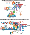
Signalling at unperturbed and perturbed replication forks. In these simplified RF models, ATR/MEC1 and CHK1/RAD53 initiate signalling pathways at RFs prior to and after passing damaged DNA resulting in stalled leading strand synthesis in the case of DNA damage, which is summarised in panels (A) and (B), respectively. In panel A, during normal, unperturbed replication processes at RFs, the Okazaki fragment synthesis on the lagging strand produces a signal via ATR/MEC1 (light red) and CHK1/RAD53 (dark pink) to slow down the RF by modulating, e.g., the CMG helicase, to synchronise DNA synthesis and nucleotide synthesis. This ATR/MEC1 activity requires Pol α and DNA2 (Okazaki fragment initiation and maturation) and signals unperturbed RFs [114,116]. When Pol ε on the leading strand encounters a DNA lesion (pink star) or is exposed to nucleotide depletion, the enzyme stops DNA synthesis and disengages with or modulates the CMG complex. Then, the latter continues to unwind DNA yielding stretches of ssDNA, which are boundby RPA. These RPA–ssDNA structures recruit ATRIP and ATR (dark red) to the leading strand, in addition to ATR’s binding via TOPBP1, its main activator, to the lagging strand with Pol α and 9-1-1 as partners (panel B). Additionally, the binding of Pol ε to TOPBP1, which may occur via a binding site of Pol ε to TOPBP1 hidden when associated with the CMG complex, may enhance this DDR signalling. It is important to note that Pol α and the RNA primer synthesis are key for the initiation of replication stress signals via ATR/MEC1 and that at RFs, Pol α does not synthesise RNA primers on the leading strand [34,72,73,117,118]. The brown, pink, and red arrows indicate ATR/MEC1 and CHK1/RAD53 signalling during unperturbed and perturbed DNA replication. This model suggests that ATR signalling requires multiple key partners located on both template strands of a stalled RF. It is important to mention that human and yeast CHK1 are homologues on the sequence level, whereas human CHK2 is the orthologue of yeast RAD53, but regarding the ATR pathway during replication stress, human CHK1 and RAD53 are functionally equivalent [119]. The figure was created with Bio Render using published results [32,72,73,114,116,117,119,120,121,122,123,124].

Similar articles

Cited by

References

    1. Burgers P.M.J., Kunkel T.A. Eukaryotic DNA Replication Fork. Annu. Rev. Biochem. 2017;86:417–438. doi: 10.1146/annurev-biochem-061516-044709. - DOI - PMC - PubMed
    1. Bleichert F., Botchan M.R., Berger J.M. Mechanisms for initiating cellular DNA replication. Science. 2017;355:eaah6317. doi: 10.1126/science.aah6317. - DOI - PubMed
    1. Costa A., Diffley J.F.X. The Initiation of Eukaryotic DNA Replication. Annu. Rev. Biochem. 2022;91:107–131. doi: 10.1146/annurev-biochem-072321-110228. - DOI - PubMed
    1. Costa A., Hood I.V., Berger J.M. Mechanisms for initiating cellular DNA replication. Annu. Rev. Biochem. 2013;82:25–54. doi: 10.1146/annurev-biochem-052610-094414. - DOI - PMC - PubMed
    1. Masai H., Matsumoto S., You Z., Yoshizawa-Sugata N., Oda M. Eukaryotic chromosome DNA replication: Where, when, and how? Annu. Rev. Biochem. 2010;79:89–130. doi: 10.1146/annurev.biochem.052308.103205. - DOI - PubMed

MeSH terms

Grants and funding

This research received no external funding.