iBet uBet web content aggregator. Adding the entire web to your favor.


Link to original content: http://pubmed.ncbi.nlm.nih.gov/38139861/
Sialic Acid Mimetic Microglial Sialic Acid-Binding Immunoglobulin-like Lectin Agonism: Potential to Restore Retinal Homeostasis and Regain Visual Function in Age-Related Macular Degeneration - PubMed Skip to main page content
U.S. flag

An official website of the United States government

Dot gov

The .gov means it’s official.
Federal government websites often end in .gov or .mil. Before sharing sensitive information, make sure you’re on a federal government site.

Https

The site is secure.
The https:// ensures that you are connecting to the official website and that any information you provide is encrypted and transmitted securely.

Access keys NCBI Homepage MyNCBI Homepage Main Content Main Navigation
Review
. 2023 Dec 16;16(12):1735.
doi: 10.3390/ph16121735.

Sialic Acid Mimetic Microglial Sialic Acid-Binding Immunoglobulin-like Lectin Agonism: Potential to Restore Retinal Homeostasis and Regain Visual Function in Age-Related Macular Degeneration

Affiliations
Review

Sialic Acid Mimetic Microglial Sialic Acid-Binding Immunoglobulin-like Lectin Agonism: Potential to Restore Retinal Homeostasis and Regain Visual Function in Age-Related Macular Degeneration

Michael J Tolentino et al. Pharmaceuticals (Basel). .

Abstract

Age-related macular degeneration (AMD), a leading cause of visual loss and dysfunction worldwide, is a disease initiated by genetic polymorphisms that impair the negative regulation of complement. Proteomic investigation points to altered glycosylation and loss of Siglec-mediated glyco-immune checkpoint parainflammatory and inflammatory homeostasis as the main determinant for the vision impairing complications of macular degeneration. The effect of altered glycosylation on microglial maintained retinal para-inflammatory homeostasis and eventual recruitment and polarization of peripheral blood monocyte-derived macrophages (PBMDMs) into the retina can explain the phenotypic variability seen in this clinically heterogenous disease. Restoring glyco-immune checkpoint control with a sialic acid mimetic agonist targeting microglial/macrophage Siglecs to regain retinal para-inflammatory and inflammatory homeostasis is a promising therapeutic that could halt the progression of and improve visual function in all stages of macular degeneration.

Keywords: Siglecs; geographic atrophy; glycosylation; macrophages; macular degeneration; microglia; nanoparticles; polysialic acid; sialic acid.


Conflict of interest statement

The authors (A.K., M.A.G.) are full-time or (M.J.T.) part-time employees of Aviceda Therapeutics, the funder of this paper. A.J.T. and E.M.T. have no conflicts of interest.

Figures
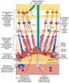
Figure 1
Figure 1
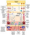
Early/intermediate-stage AMD. (1) Photo-oxidative blue light (2) oxidizes retinal lipids to produces oxidative byproducts (MDA, CEP) and reactive oxygen species, (3) which activate microglial cells to secrete cytokines. These activated macrophages are hindered from becoming phagocytic due to the (4) upregulation of ST6 that produces (5) sTN on the surface of retinal cells and drusen that agonizes Siglecs to prevent phagocytosis of drusen. (6) Reactive oxygen species also cause photoreceptors to secrete CX3CL1, which promotes migration of microglia and macrophages to the retina. (7) Oxidized phospholipids stimulate RPE cells to produce MCP—1 (8), which recruits peripheral blood monocytes (PBMCs) to areas of phospholipid oxidation. (9) Upregulation of FUT5 on PBMCs localizes monocytes to areas that are secreting chemokines such as CX3CL1 that upregulate e-selectin on vascular endothelium. (10) Monocytes are found between RPE cells and Bruch’s membrane. (11) Microglial cells and macrophages are also found to be pigment-ladened in this sub-RPE space and at the RPE cell layer, which appears as RPE pigment clumping. (12) The overactivation of complement caused by the polymorphism-induced impaired function of CFH will produce complement pathway metabolites that accumulate in drusen and can activate microglial cells.
Figure 2
Figure 2
Microglial parainflammatory regulation: Microglia remove toxic metabolites, apoptotic cells, and oxidative debris while maintaining the health of photoreceptors and retinal cells in a process called parainflammation. (1) Parainflammation is initiated by pattern recognition receptors found on (2) M0 microglia such as TLR, TREM2, CX3CR1, NLRP3, and CD36, which use these receptors to bind and sense toxins, apoptotic cells, damage, and pathogen-associated molecular patterns. Once microglia are activated, they polarize to the (3) M1 like state, (4) secrete proinflammatory cytokines, and (5) migrate to the areas of stressed RPE cells and photoreceptors. (6) Upon activation, neuropilin-2-PSA and E-selectin ligand 1-PSA are secreted by these activated microglia. (6) The PSA on these glycoproteins binds Siglecs on activated microglial cells (7) to polarize them into the M2c healing state (8), which releases growth factors to protect and regenerate the stressed photoreceptors and RPE cells. (9) This homeostatic maintenance function of microglial parainflammation, if not adequately modulated with sialic acid checkpoint regulation, will result in recruitment of peripheral blood monocytes and AMD disease progression. (10) A PSA-nanoparticle (PSA-NP) mimics E-selectin ligand 1 PSA and Neuropilin 2-PSA to (7) polarize M1 activated microglia into the M2c healing state.
Figure 3
Figure 3
Geographic atrophy: (1) Progressive accumulation of oxidative byproducts (CEP, MDA) and (2) chronic overactivated complement pathway chronically activate microglia (3), which secrete neuraminidase and desialylate photoreceptors and RPE cells. (4) This loss of sialylation prevents restoration of homeostasis. (5) Chronically overactivated phagocytosis and inflammation recruits peripheral blood macrophages by upregulation of the fucosyltransferase FUT5 that produces Lewis X glycosylation on monocytes to bind E-selectin and (6) localize monocytes to sites of inflammation (7), allowing for diapedesis of the monocyte across the blood–retinal barrier. (8) These monocytes polarize to M1 macrophages when they enter the retina and are not able to clear substances like lipofuscin and other undegradable substances. (9) The macrophages form multinucleated giant cells because they phagocytose structures that are undegradable. (10) Since there is no sialic acid or polysialic acid to polarize to the healing M2c state, the macrophages are unchecked and result in elimination (11) first of the RPE cells then the (12) photoreceptors. The unchecked macrophages are the main determinant of growth of geographic atrophy.
Figure 4
Figure 4
Macrophage polarization determines AMD phenotype. The plasticity and different polarization states correlate with the clinical picture seen in late-stage macular degeneration. In geographic atrophy, RPE cells and photoreceptors are phagocytosed as a function of the M1 polarized phagocytic macrophages. Exudative AMD is neovascularization produced by overexpression of VEGF, the main cytokine secreted by the M2d polarization state. The sequelae of exudative AMD untreated with anti-VEGF therapies is a disciform fibrotic scar in the control of the pro-fibrotic M2 A, B state. With appropriate sialic acid signaling such as PSA, all polarization states can transform into the healing M2c state.
Figure 5
Figure 5
PSA-NP can halt GA and restore vision. PSA-NP can multivalently bind Siglecs on polarized macrophages and microglia, transforming them into the M2c healing state. These M2c healing macrophages/microglia will polarize M1 inflammatory, M1 phagocytic, M2D angiogenic, and M2a, b fibrotic M2c neuroprotective and BDNF- and GDNF-secreting macrophages. This in turn will rescue photoreceptors and RPE cells and restore the homeostatic parainflammatory function of microglia, potentially halting GA progression, reducing lipofuscin and improving visual function and vision.

Similar articles

Cited by

References

    1. Taylor D.J., Hobby A.E., Binns A.M., Crabb D.P. How does age-related macular degeneration affect real-world visual ability and quality of life? A systematic review. BMJ Open. 2016;6:e011504. doi: 10.1136/bmjopen-2016-011504. - DOI - PMC - PubMed
    1. Scott A.W., Bressler N.M., Ffolkes S., Wittenborn J.S., Jorkasky J. Public Attitudes About Eye and Vision Health. JAMA Ophthalmol. 2016;134:1111–1118. doi: 10.1001/jamaophthalmol.2016.2627. - DOI - PubMed
    1. Keenan T.D.L., Cukras C.A., Chew E.Y. Age-Related Macular Degeneration: Epidemiology and Clinical Aspects. Adv. Exp. Med. Biol. 2021;1256:1–31. doi: 10.1007/978-3-030-66014-7_1. - DOI - PubMed
    1. Izekenova A.K., Kumar A.B., Abikulova A.K., Izekenova A.K. Trends in ageing of the population and the life expectancy after retirement: A comparative country-based analysis. J. Res. Med. Sci. 2015;20:250–252. - PMC - PubMed
    1. Rein D.B., Wittenborn J.S., Burke-Conte Z., Gulia R., Robalik T., Ehrlich J.R., Lundeen E.A., Flaxman A.D. Prevalence of Age-Related Macular Degeneration in the US in 2019. JAMA Ophthalmol. 2022;140:1202–1208. doi: 10.1001/jamaophthalmol.2022.4401. - DOI - PMC - PubMed

LinkOut - more resources