iBet uBet web content aggregator. Adding the entire web to your favor.


Link to original content: http://pubmed.ncbi.nlm.nih.gov/37279536/
The pyrenoid: the eukaryotic CO2-concentrating organelle - PubMed Skip to main page content
U.S. flag

An official website of the United States government

Dot gov

The .gov means it’s official.
Federal government websites often end in .gov or .mil. Before sharing sensitive information, make sure you’re on a federal government site.

Https

The site is secure.
The https:// ensures that you are connecting to the official website and that any information you provide is encrypted and transmitted securely.

Access keys NCBI Homepage MyNCBI Homepage Main Content Main Navigation
. 2023 Sep 1;35(9):3236-3259.
doi: 10.1093/plcell/koad157.

The pyrenoid: the eukaryotic CO2-concentrating organelle

Affiliations

The pyrenoid: the eukaryotic CO2-concentrating organelle

Shan He et al. Plant Cell. .

Abstract

The pyrenoid is a phase-separated organelle that enhances photosynthetic carbon assimilation in most eukaryotic algae and the land plant hornwort lineage. Pyrenoids mediate approximately one-third of global CO2 fixation, and engineering a pyrenoid into C3 crops is predicted to boost CO2 uptake and increase yields. Pyrenoids enhance the activity of the CO2-fixing enzyme Rubisco by supplying it with concentrated CO2. All pyrenoids have a dense matrix of Rubisco associated with photosynthetic thylakoid membranes that are thought to supply concentrated CO2. Many pyrenoids are also surrounded by polysaccharide structures that may slow CO2 leakage. Phylogenetic analysis and pyrenoid morphological diversity support a convergent evolutionary origin for pyrenoids. Most of the molecular understanding of pyrenoids comes from the model green alga Chlamydomonas (Chlamydomonas reinhardtii). The Chlamydomonas pyrenoid exhibits multiple liquid-like behaviors, including internal mixing, division by fission, and dissolution and condensation in response to environmental cues and during the cell cycle. Pyrenoid assembly and function are induced by CO2 availability and light, and although transcriptional regulators have been identified, posttranslational regulation remains to be characterized. Here, we summarize the current knowledge of pyrenoid function, structure, components, and dynamic regulation in Chlamydomonas and extrapolate to pyrenoids in other species.


Conflict of interest statement

Conflict of interest statement. None declared.

Figures

Figure 1.
Figure 1.
Structure of the Chlamydomonas pyrenoid. A) The Chlamydomonas pyrenoid is visible by light microscopy. Scale bar, 1 μm. B) The Chlamydomonas pyrenoid is composed of three major compartments: the Rubisco matrix, thylakoid tubules, and starch sheath. Scale bar, 200 nm. C) Two-dimensional and D) Three-dimensional models of the pyrenoid showing the major compartments and protein peripheral structures. See Table 1 for a list of the known protein components of each structure. (The circled numbers indicating the sub-pyrenoid localizations in panel C are coordinated with the circled numbers in Table 1).
Figure 2.
Figure 2.
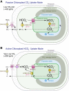
Operating principles of the pyrenoid-based CCM. The Chlamydomonas CO2-concentrating mechanism is shown; the basic principles are likely to apply in other species, although due to convergent evolution, the specific proteins that mediate some of the reactions may be phylogenetically unrelated to those in Chlamydomonas. Mutant phenotypes and biophysical modeling (Fei et al. 2022) support the existence of two operating modes of a pyrenoid-based CO2-concentrating mechanism, which differ based on how HCO3 is accumulated in the chloroplast stroma. A) The first mode uses a passive chloroplast CO2 uptake strategy, where CO2 passively diffuses across the chloroplast envelope into the stroma and is converted into HCO3 by the LCIB/LCIC carbonic anhydrase complex. This strategy is used under low CO2 (ambient air levels of external CO2). B) The second mode uses an active chloroplast HCO3 uptake strategy, which relies on active pumping of HCO3 into the chloroplast. This strategy is used under very low CO2.
Figure 3.
Figure 3.
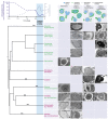
Pyrenoids appear to have convergently evolved in response to declining atmospheric CO2 levels. Approximate CO2 and O2 concentrations over time (Berner 2006; Whitney et al. 2011) are correlated with the phylogenetic tree of photosynthetic eukaryotes below (Strassert et al. 2021). Branch points correlate with the approximate timing of the divergence of different groups (see Strassert et al. 2021 and Bowles et al. 2022 for discussions on uncertainties regarding branch points). Asterisks denote the approximate timing of the acquisition of plastids through primary (*) or secondary (**) endosymbiosis (Jackson et al. 2018; Strassert et al. 2021). The blue shade highlights the proposed range for the timing of CCM evolution in different photosynthetic species (Villarreal and Renner 2012; Meyer et al. 2020a). Green and red lineages are denoted as green or purple text, respectively (most dinoflagellates have red plastids with the exception of Lepidodinium sp., which have green plastids) (Kamikawa et al. 2015). Representative electron micrographs of pyrenoids are shown below cartoons of four general pyrenoid types, displaying the wide variety of morphologies observed in each algal lineage and the hornworts. Roman numerals on the electron micrographs denote references to their original publications as follows: (I) Kusel-Fetzmann and Weidinger 2008; (II) Nudelman et al. 2006; (III) Zhang et al. 2008; (IV) van Baren et al. 2016; (V) Borowitzka 2018; (VI) Goudet et al. 2020; (VII) Mikhailyuk et al. 2014; (VIII) Duff et al. 2007; (IX) Hall and Claus 1963; (X) Nelson and Ryan 1988; (XI) Ford 1984; (XII) Laza-Martínez et al. 2012; (XIII) Clay and Kugrens 1999; (XIV) Shiratori et al. 2017; (XV) Ota et al. 2007; (XVI) Schnepf and ElbräChter 1999; (XVII) Kowallik 1969; (XVIII) Hansen and Daugbjerg 2009; (XIX) Decelle et al. 2021; (XX) Bedoshvili et al. 2009; (XXI) Bedoshvili and Likhoshway 2012; (XXII) Buma et al. 2000; (XXIII) Fresnel and Probert 2005. This figure was created with BioRender.
Figure 4.
Figure 4.
A common Rubisco-binding motif mediates the assembly of the major compartments of the pyrenoid. A) In Chlamydomonas, many pyrenoid-localized proteins contain at least one Rubisco-binding motif. B) The Rubisco-binding motif mediates the assembly of the three pyrenoid sub-compartments. The motifs on EPYC1 link Rubisco to form the pyrenoid matrix (He et al. 2020). The motifs on the tubule-localized transmembrane proteins RBMP1 and RBMP2 are proposed to connect the Rubisco to the tubules, and the motifs on the putative starch-binding proteins SAGA1 and SAGA2 are proposed to mediate interactions between the matrix and the surrounding starch sheath. A Rubisco-binding motif was also shown to be necessary and sufficient to target a nascent protein to the pyrenoid (Meyer et al. 2020b). C) A model illustrating how EPYC1 (red) clusters Rubisco (blue) in the pyrenoid matrix. D) The Rubisco-binding motif of EPYC1 (red) binds to the Rubisco small subunit (dark blue) (He et al. 2020); other Rubisco-binding motifs in Chlamydomonas are expected to bind to the same site. E) The motif binds between two alpha-helices of the Rubisco small subunit.
Figure 5.
Figure 5.
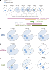
The Chlamydomonas pyrenoid exhibits liquid-like behavior during cell divisions. A) Diagram depicting the timeline and morphology of a typical cell division with pyrenoid fission in Chlamydomonas (adapted from Freeman Rosenzweig et al. 2017). The time point t = 0 is the moment the chloroplast division furrow passes between the daughter pyrenoids. A portion of the pyrenoid matrix disperses into the chloroplast stroma during the division of the pyrenoid. The approximate timing and duration of key events are shown below the timeline. B) Diagram depicting the “bridge” of matrix during pyrenoid fission. C) Diagram depicting the transient appearance of small puncta of pyrenoid matrix throughout the stroma during dispersal of the matrix in some dividing cells. D) Diagram depicting the de novo formation of a daughter pyrenoid when pyrenoid fission fails. The lower daughter cell inherits the entire pyrenoid of the mother cell. The upper cell shows de novo pyrenoid formation with the appearance of one or more fluorescent puncta growing or coalescing into one pyrenoid (observed in wild-type cells expressing either EPYC1-Venus or Rubisco-Venus).

Similar articles

Cited by

References

    1. Adler L, Diaz-Ramos A, Mao Y, Pukacz KR, Fei C, McCormick AJ. New horizons for building pyrenoid-based CO2-concentrating mechanisms in plants to improve yields. Plant Physiol. 2022:190(3):1609–1627. 10.1093/plphys/kiac373 - DOI - PMC - PubMed
    1. Anbar AD, Duan Y, Lyons TW, Arnoled GL, Kendall B. A whiff of oxygen before the great oxidation event? Science. 2007:317(5846):1903–1906. 10.1126/science.1140325 - DOI - PubMed
    1. Ang WSL, How JA, How JB, Mueller-Cajar O. The stickers and spacers of Rubisco condensation: assembling the heartpiece of biophysical CO2 concentrating mechanisms. J Exp Bot. 2022:74(2):612–626. 10.1093/jxb/erac321 - DOI - PubMed
    1. Atkinson N, Mao Y, Chan KX, McCormick AJ. Condensation of Rubisco into a proto-pyrenoid in higher plant chloroplasts. Nat Commun. 2020:11(1):6303. 10.1038/s41467-020-20132-0 - DOI - PMC - PubMed
    1. Atkinson N, Velanis CN, Wunder T, Clarke DJ, Mueller-Cajar O, McCormick AJ. The pyrenoidal linker protein EPYC1 phase separates with hybrid Arabidopsis-Chlamydomonas Rubisco through interactions with the algal Rubisco small subunit. J Exp Bot. 2019:70(19):5271–5285. 10.1093/jxb/erz275 - DOI - PMC - PubMed

Publication types

MeSH terms

Substances