iBet uBet web content aggregator. Adding the entire web to your favor.


Link to original content: http://pubmed.ncbi.nlm.nih.gov/36538378/
Loss of LGR4/GPR48 causes severe neonatal salt wasting due to disrupted WNT signaling altering adrenal zonation - PubMed Skip to main page content
U.S. flag

An official website of the United States government

Dot gov

The .gov means it’s official.
Federal government websites often end in .gov or .mil. Before sharing sensitive information, make sure you’re on a federal government site.

Https

The site is secure.
The https:// ensures that you are connecting to the official website and that any information you provide is encrypted and transmitted securely.

Access keys NCBI Homepage MyNCBI Homepage Main Content Main Navigation
. 2023 Feb 15;133(4):e164915.
doi: 10.1172/JCI164915.

Loss of LGR4/GPR48 causes severe neonatal salt wasting due to disrupted WNT signaling altering adrenal zonation

Affiliations

Loss of LGR4/GPR48 causes severe neonatal salt wasting due to disrupted WNT signaling altering adrenal zonation

Cécily Lucas et al. J Clin Invest. .

Abstract

Disorders of isolated mineralocorticoid deficiency, which cause potentially life-threatening salt-wasting crisis early in life, have been associated with gene variants of aldosterone biosynthesis or resistance; however, in some patients no such variants are found. WNT/β-catenin signaling is crucial for differentiation and maintenance of the aldosterone-producing adrenal zona glomerulosa (zG). Herein, we describe a highly consanguineous family with multiple perinatal deaths and infants presenting at birth with failure to thrive, severe salt-wasting crises associated with isolated hypoaldosteronism, nail anomalies, short stature, and deafness. Whole exome sequencing revealed a homozygous splice variant in the R-SPONDIN receptor LGR4 gene (c.618-1G>C) regulating WNT signaling. The resulting transcripts affected protein function and stability and resulted in loss of Wnt/β-catenin signaling in vitro. The impact of LGR4 inactivation was analyzed by adrenal cortex-specific ablation of Lgr4, using Lgr4fl/fl mice mated with Sf1:Cre mice. Inactivation of Lgr4 within the adrenal cortex in the mouse model caused decreased WNT signaling, aberrant zonation with deficient zG, and reduced aldosterone production. Thus, human LGR4 mutations establish a direct link between LGR4 inactivation and decreased canonical WNT signaling, which results in abnormal zG differentiation and endocrine function. Therefore, variants in WNT signaling and its regulators should systematically be considered in familial hyperreninemic hypoaldosteronism.

Keywords: Endocrinology; Genetic diseases; Molecular biology; Mouse models.


Figures
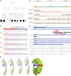
Figure 1
Figure 1. Genetic and structural characterization of a human LGR4 mutation identified in a highly consanguineous family.
(A) Family pedigree showing first-degree consanguinity and multiple affected individuals. Squares: male; circles: female; diamonds: unknown sex; triangle: miscarriage. Black symbols represent affected and white symbols represent unaffected individuals. Numbers in symbols represent multiple individuals. The index patient is indicated by a black arrowhead. Asterisks indicate individuals for whom DNA was sequenced. (B) Partial chromatograms showing the LGR4 mutation NM_018490.5:c.618-1 G>C. Intron 5 (red) and exon 6 (blue) are highlighted. The proband is homozygous, parents and 1 brother are heterozygous, and 1 brother had the WT sequence. (C) LGR4 mRNA analysis of proband fibroblasts. The first track represents patient mRNA after reverse transcription, indicating presence of 2 transcripts. The 2 transcripts were separated by cloning and sequenced (2 middle tracks). The bottom track shows cDNA of control fibroblasts. The scheme above shows normal splicing in dark lines and the effect of the mutation on splicing in blue dotted lines. (D) Amino acid sequence of extracellular domain of human LGR4 that binds to RSPOs. Amino acids found deleted in the patient are located in LRR7 (–8 AA) and LRR7/8 (–24 AA) coded by exon 6 of LGR4. (E) Structure of LGR4 and its interaction with RSPOs. (i) Structure of human LGR4 extracellular domain in complex with part of RSPO1 (PDB 4KT1). Amino acids coded by exon 6 are depicted in red. (ii) Human LGR4-RSPO3 complex. RSPO3 shares high structural similarity with RSPO1 and binds to LRR7 and LRR8 of LGR4. (iii and iv) Patient LGR4, missing 8 or 24 amino acids. Several critical hydrogen-bonding residues in LGR4 are missing due to mutations, causing weaker interaction and binding of RSPO3 to mutant LGR4 proteins. (v) A surface view of the LGR4-RSPO3 complex, showing close interaction points. Exon 6–encoded residues are shown in red.
Figure 2
Figure 2. Protein expression and functional testing of Wnt/β-catenin signaling of the 2 LGR4 variants.
Human fibroblasts and HEK cells were used. (A) Western blot analysis for LGR4 protein expression in patient and control fibroblasts. A representative blot of 3 independent experiments is shown. β-Actin was used as a loading control. The molecular weight (kDa) of a protein standard is given. M, marker; Co, control fibroblasts; Pat, patient fibroblasts. (B) RSPO1-activated, LGR4-mediated Wnt signaling in HEK293 cells. Cells were transfected with WT and mutant LGR4 nt-24 and nt-72 plasmids (including a mock control) and reporter vectors TOP-Flash and Renilla. Signaling was stimulated by RSPO1 and assessed by the Dual-Luciferase assay (Promega). Results are expressed as relative LUC activities (RLU). Mean ± SD of 3 independent experiments is shown. *P < 0.01, Student’s t test. (C and D) Interaction of RSPO1 with membrane-localized WT and variant LGR4. HEK293 cells were transfected with HA-tagged LGR4 plasmids (pcDNA3 LGR4wt, nt-24 bp, nt-72 bp) and incubated with conditioned RSPO1-GFP SN medium (previously produced in HEK cells transfected with pSpark- RSPO1-GFP). Cells were fixed with Carnoy’s solution. Staining was performed with antibody anti–HA-Tag (green) first and antibody anti-mouse Alexa Fluor 594 (red) second. Immunofluorescent microscopy was used to detect the cellular distribution of the tagged proteins as well as their colocalization (Zeiss LSM 710). Three independent experiments were analyzed. Representative images of confocal analysis at original magnification, ×40 are shown for WT and variants of LGR4. Scale bars: 5 μm (top 2 images in middle column and right); 10 μm (left and bottom image in the middle column). Quantification of colocalized LGR4 and RSPO1 was performed by Imaris (Bitplane AG).
Figure 3
Figure 3. Lgr4 ablation disrupts the Wnt/β-catenin signaling pathway, resulting in adrenal hypoplasia and aberrant zonal differentiation.
(A) RT-qPCR analysis of mRNA encoding Wnt/β-catenin signaling pathway–associated genes. (B) Immunohistochemical detection of β-catenin and Lef1. (C) LEF1+ cell index, defined as the percentage of LEF1+ cells over the total number of cortical cells. (D) Adrenal weight. (E) H&E staining of WT and Lgr4cKO adrenals. (F) Number of cortical cells per 500 μm2 of the cortex. (G) Coimmunostaining for Akr1b7 and Dab2 in WT and Lgr4cKO adrenals. (H) RT-qPCR analysis of mRNA encoding zone-specific markers (Akr1b7, Dab2, and Hsd3b6). All analyses were conducted in 5-week-old WT and Lgr4cKO female mice. zF, zona fasciculata; zG, zona glomerulosa; M, medulla; Co, cortex. Scale bars: 50 μm. Data are shown as the mean ± SEM. Numbers of individual samples analyzed are indicated within the bars. Statistical analyses in A, C, D, F, and H were conducted using Mann-Whitney tests in GraphPad Prism 9. *P < 0.05, ***P < 0.001, ****P < 0.0001.
Figure 4
Figure 4. Lgr4 ablation inhibits zona glomerulosa differentiation, resulting in primary hypoaldosteronism.
(A) Aldosterone plasma concentration and (B) renin activity in 5-week-old WT and Lgr4cKO female mice. (C) Immunohistochemical detection of CYP11B2. Scale bar: 50 μm. zF, zona fasciculata; zG, zona glomerulosa; M, medulla. (D) Number of CYP11B2+ cells per adrenal section. (E) Corticosterone and (F) plasma ACTH concentration. (G) RT-qPCR analysis of mRNA encoding steroidogenesis-related genes. All analyses were conducted in 5 weeks WT and Lgr4cKO female mice. Data are shown as the mean ± SEM. Numbers of individual samples analyzed are indicated within the bars. Statistical analyses in A, B, and DG were conducted using Mann-Whitney tests in GraphPad Prism 9. *P < 0.05, **P < 0.01.

Similar articles

Cited by

References

    1. Miller WL, et al. In: Sperling MA, ed. Sperling - Pediatric Endocrinology. Elsevier; 2020.
    1. Holtback U, Aperia AC. Molecular determinants of sodium and water balance during early human development. Semin Neonatol. 2003;8(4):291–299. doi: 10.1016/S1084-2756(03)00042-3. - DOI - PubMed
    1. Martinerie L, et al. Physiological partial aldosterone resistance in human newborns. Pediatr Res. 2009;66(3):323–328. doi: 10.1203/PDR.0b013e3181b1bbec. - DOI - PMC - PubMed
    1. Rosler A. The natural history of salt-wasting disorders of adrenal and renal origin. J Clin Endocrinol Metab. 1984;59(4):689–700. doi: 10.1210/jcem-59-4-689. - DOI - PubMed
    1. Fu Y, Vallon V. Mineralocorticoid-induced sodium appetite and renal salt retention: evidence for common signaling and effector mechanisms. Nephron Physiol. 2014;128(1-2):8–16. doi: 10.1159/000368264. - DOI - PMC - PubMed

Publication types

Grants and funding

Pierre Val is supported by a program grant (Labellisation) by the “Ligue Nationale Contre le Cancer” 2021-2025.