iBet uBet web content aggregator. Adding the entire web to your favor.


Link to original content: http://pubmed.ncbi.nlm.nih.gov/35856560/
Multiple Photolyases Protect the Marine Cyanobacterium Synechococcus from Ultraviolet Radiation - PubMed Skip to main page content
U.S. flag

An official website of the United States government

Dot gov

The .gov means it’s official.
Federal government websites often end in .gov or .mil. Before sharing sensitive information, make sure you’re on a federal government site.

Https

The site is secure.
The https:// ensures that you are connecting to the official website and that any information you provide is encrypted and transmitted securely.

Access keys NCBI Homepage MyNCBI Homepage Main Content Main Navigation
. 2022 Aug 30;13(4):e0151122.
doi: 10.1128/mbio.01511-22. Epub 2022 Jul 20.

Multiple Photolyases Protect the Marine Cyanobacterium Synechococcus from Ultraviolet Radiation

Affiliations

Multiple Photolyases Protect the Marine Cyanobacterium Synechococcus from Ultraviolet Radiation

Allissa M Haney et al. mBio. .

Abstract

Marine cyanobacteria depend on light for photosynthesis, restricting their growth to the photic zone. The upper part of this layer is exposed to strong UV radiation (UVR), a DNA mutagen that can harm these microorganisms. To thrive in UVR-rich waters, marine cyanobacteria employ photoprotection strategies that are still not well defined. Among these are photolyases, light-activated enzymes that repair DNA dimers generated by UVR. Our analysis of genomes of 81 strains of Synechococcus, Cyanobium, and Prochlorococcus isolated from the world's oceans shows that they possess up to five genes encoding different members of the photolyase/cryptochrome family, including a photolyase with a novel domain arrangement encoded by either one or two separate genes. We disrupted the putative photolyase-encoding genes in Synechococcus sp. strain RS9916 and discovered that each gene contributes to the overall capacity of this organism to survive UVR. Additionally, each conferred increased survival after UVR exposure when transformed into Escherichia coli lacking its photolyase and SOS response. Our results provide the first evidence that this large set of photolyases endows Synechococcus with UVR resistance that is far superior to that of E. coli, but that, unlike for E. coli, these photolyases provide Synechococcus with the vast majority of its UVR tolerance. IMPORTANCE Cells use DNA photolyases to protect their DNA from the damaging effects of UV radiation. Marine cyanobacteria possess many genes that appear to encode photolyases, but the function of the proteins encoded by these genes is unclear. The study uses comparative genomics and molecular genetic approaches to describe and characterize the roles of these proteins in DNA damage repair in the marine cyanobacterium Synechococcus. This study identifies the important role of DNA photolyases in DNA repair for these cells and describes a previously undescribed structural class of DNA of these enzymes.

Keywords: DNA photolyase; Synechococcus; UV light; cyanobacteria; marine microbiology.


Conflict of interest statement

The authors declare no conflict of interest.

Figures

FIG 1
FIG 1
Structural domains and bound chromophores for the eight members of the cryptochrome/photolyase family found in marine and brackish picocyanobacteria and illustration of the phr4/phr5 junction. (A) Diagrams show the positions of the different protein domains, as predicted from InterProScan (108) in representative sequences of the eight Cyanorak v2.1 clusters of likely orthologous genes (CLOGs), indicated between brackets after the protein names. Sequences shown here are from Synechococcus sp. RS9916 for Phr1-5, from Cyanobium sp. NS01 for Phr6, from Prochlorococcus sp. MIT9302 for Phr7, and from C. gracile PCC 6307 for Phr8. By analogy with the freshwater Synechococcus sp. (formerly Anacystis nidulans) strain PCC 6301 (109), we assume that an 8-hydroxy-5-deazaflavin (8-HDF), represented as a purple diamond, is bound to the DNA photolyase domain (InterPro accession no. IPR036155; 109) of Phr3. The chromophores bound to the other DNA photolyase domains shown, and to the photolyase PhrB-like domain (IPR007357), have not yet been identified and are denoted by a gray diamond. Additionally, blue diamonds indicate that the amino acids involved in flavin binding to the FAD binding domain (IPR036134), identified in Synechococcus sp. PCC 6301 Syc1392_c (Y228, T240, S241, L243, S244, W280, R287, T346, N349, D380, D382, A385, and N386), are conserved in the FAD domains of all CLOG members (e.g., Y247, T259, S260, L262, S263, W299, R306, W365, N368, D399, D401, A404, and N405 in 9916 Phr3). Finally, red stars indicate that the corresponding FAD domains contain the residues necessary to bind an Fe-S complex (e.g., in RS9916 Phr4: C169, C254, C257, and C263), as found in the (6-4) photolyase from A. tumefaciens (46). (B) DNA and translated protein sequences from the genomic region spanning phr4 and phr5 in 9916. The DNA sequence encoding the stop codon (asterisk) of phr4 is in green, while the start codon of phr5 is in red.
FIG 2
FIG 2
Maximum likelihood phylogenetic tree of the cryptochrome/photolyase family based on the FAD domain. Marine picocyanobacteria sequence members retrieved from 81 nonredundant genomes from Cyanorak v2.1 (16) are shown in colors, with monophyletic groups within each protein member being collapsed as colored triangles (the noncollapsed tree is shown in Fig. S1). Outgroup sequences are shown in black. Picocyanobacterial sequence names include the abbreviation of the genus (Pro, Prochlorococcus; Syn, Synechococcus; Cya, Cyanobium), strain name, and subcluster sensu Herdman et al. (110), as well as the Cyanorak CLOG number and the proposed protein designation as in Fig. 1 and Table S1. Plain gray circles on branches of the tree correspond to maximum likelihood bootstrap values ranging from 70 to 100% (lower values have been omitted). The red star indicates the members that possess the four conserved cysteine residues necessary to bind an Fe-S cluster, as found, for example, in the (6-4) photolyase from A. tumefaciens (46) and V. cholerae O395 (Table S2). Note that Phr5 is not shown since it does not possess a FAD domain.
FIG 3
FIG 3
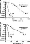
Percent survival rates of two bacterial species after UV-B and UV-C treatment. (A and B) Percent survival rates of E. coli (circles) and marine Synechococcus RS9916 (squares) after various doses of (A) UV-B and (B) UV-C radiation followed by continuous white light (WL) exposure. For each species, the number of cells on plates not treated with UV radiation but exposed to continuous WL were used to establish the 100% value. Error bars denote the standard deviation of at least three independent replicates.
FIG 4
FIG 4
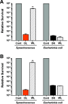
Light-dependent survival of Synechococcus 9916 and E. coli cells after UVR treatment. (A and B) After treatment with (A) 1,728 J m−2 of UV-B or (B) 212 J m−2 of UV-C, 9916 cells (left) were placed in either orange light (OL) or white light (WL), while E. coli cells (right) were placed in either the dark (DK) or white light (WL). After 1 h, a dilution series of cells were plated and grown under the same light conditions, and colony numbers were counted. For both organisms, a no-UV treatment control was included (Cont), and the values were normalized to 1. Error bars are the standard deviation (SD) of at least three replicates. **, P < 0.01.
FIG 5
FIG 5
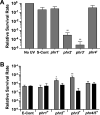
Contribution of putative photolyase-encoding genes to percent survival rates in Synechococcus and E. coli after UV-B treatment. (A) Synechococcus 9916 control cells were either not exposed to UV-B (No UV) or exposed to 1,728 J m−2 of UV-B followed by WL treatment (S-Cont), while four mutants containing insertions in putative photolyase-encoding genes (phr1, phr2, phr3, phr4) were given the equivalent UV-B and WL treatment. Values obtained for the no-UV control cells were set 1. **, P < 0.01, compared to control cells. (B) An E. coli mutant lacking photolyase activity and the SOS response was transformed with a vector only and either not exposed to UV-B or exposed to 154 J m−2 of UV-B (E-Cont) followed either by WL (gray bar) or dark (black bar) treatment. The same E. coli mutant was transformed with the same vector carrying either the Synechococcus 9916 phr1, phr2, phr3, or phr4/phr5 gene and exposed to an equivalent dose of UV-B followed either by WL (gray bar) or dark (black bar) treatment. The data for the vector-only transformed E. coli cells that were not treated with UV-B were set to a value of 1 and are not shown in panel B. Error bars are the SD of at least three replicates. *, P < 0.05; **, P < 0.01, compared to light-treated control cells.
FIG 6
FIG 6
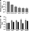
Contribution of putative photolyase-encoding genes to percent survival rates in Synechococcus and E. coli after UV-C treatment. (A) Synechococcus 9916 control cells were either not exposed to UV-C (No UV) or exposed to 212 J m−2 of UV-C followed by WL treatment (S-Cont), while four mutants containing insertions in putative photolyase-encoding genes (phr1, phr2, phr3, phr4) were given the equivalent UV-C and WL treatment. Values obtained for the no-UV control cells were set to a value of 1. **, P < 0.01, compared to control cells. (B) An E. coli mutant lacking photolyase activity and the SOS response was transformed with a vector only and either not exposed to UV-C or exposed to 24 J m−2 of UV-C (E-Cont) followed either by WL (gray bar) or dark (black bar) treatment. The same E. coli mutant was transformed with the same vector carrying either the Synechococcus 9916 phr1, phr2, phr3, or phr4/5 gene and exposed to an equivalent dose of UV-C followed either by WL (gray bar) or dark (black bar) treatment. The data for the vector-only transformed E. coli cells that were not treated with UV-C were set to a value of 1 and are not shown in panel B. Error bars are the SD of at least three replicates. *, P < 0.05; **, P < 0.01, compared to light-treated control cells.

Similar articles

Cited by

References

    1. Behrenfeld MJ, O’Malley RT, Siegel DA, McClain CR, Sarmiento JL, Feldman GC, Milligan AJ, Falkowski PG, Letelier RM, Boss ES. 2006. Climate-driven trends in contemporary ocean productivity. Nature 444:752–755. doi:10.1038/nature05317. - DOI - PubMed
    1. Llabres M, Agusti S. 2006. Picophytoplankton cell death induced by UV radiation: evidence for oceanic Atlantic communities. Limnol Oceanogr 51:21–29. doi:10.4319/lo.2006.51.1.0021. - DOI
    1. Agusti S, Llabres M. 2007. Solar radiation-induced mortality of marine pico-phytoplankton in the oligotrophic ocean. Photochem Photobiol 83:793–801. doi:10.1111/j.1751-1097.2007.00144.x. - DOI - PubMed
    1. Hader DP, Kumar HD, Smith RC, Worrest RC. 2007. Effects of solar UV radiation on aquatic ecosystems and interactions with climate change. Photochem Photobiol Sci 6:267–285. doi:10.1039/b700020k. - DOI - PubMed
    1. Llabres M, Agusti S, Alonso-Laita P, Herndl GJ. 2010. Synechococcus and Prochlorococcus cell death induced by UV radiation and the penetration of lethal UVR in the Mediterranean Sea. Mar Ecol Prog Ser 399:27–37. doi:10.3354/meps08332. - DOI

Publication types

LinkOut - more resources