iBet uBet web content aggregator. Adding the entire web to your favor.


Link to original content: http://pubmed.ncbi.nlm.nih.gov/35659639/
Regulation of Rostral Nucleus of the Solitary Tract Responses to Afferent Input by A-type K+ Current - PubMed Skip to main page content
U.S. flag

An official website of the United States government

Dot gov

The .gov means it’s official.
Federal government websites often end in .gov or .mil. Before sharing sensitive information, make sure you’re on a federal government site.

Https

The site is secure.
The https:// ensures that you are connecting to the official website and that any information you provide is encrypted and transmitted securely.

Access keys NCBI Homepage MyNCBI Homepage Main Content Main Navigation
. 2022 Jul 15:495:115-125.
doi: 10.1016/j.neuroscience.2022.05.036. Epub 2022 Jun 2.

Regulation of Rostral Nucleus of the Solitary Tract Responses to Afferent Input by A-type K+ Current

Affiliations

Regulation of Rostral Nucleus of the Solitary Tract Responses to Afferent Input by A-type K+ Current

Z Chen et al. Neuroscience. .

Abstract

Responses in the rostral (gustatory) nucleus of the solitary tract (rNST) are modified by synaptic interactions within the nucleus and the constitutive membrane properties of the neurons themselves. The potassium current IA is one potential source of modulation. In the caudal NST, projection neurons with IA show lower fidelity to afferent stimulation compared to cells without. We explored the role of an A-type K+ current (IA) in modulating the response to afferent stimulation and GABA-mediated inhibition in the rNST using whole cell patch clamp recording in transgenic mice that expressed channelrhodopsin (ChR2 H134R) in GABAergic neurons. The presence of IA was determined in current clamp and the response to electrical stimulation of afferent fibers in the solitary tract was assessed before and after treatment with the specific Kv4 channel blocker AmmTX3. Blocking IA significantly increased the response to afferent stimulation by 53%. Using dynamic clamp to create a synthetic IA conductance, we demonstrated a significant 14% decrease in responsiveness to afferent stimulation in cells lacking IA. Because IA reduced excitability and is hyperpolarization-sensitive, we examined whether IA contributed to the inhibition resulting from optogenetic release of GABA. Although blocking IA decreased the percent suppression induced by GABA, this effect was attributable to the increased responsiveness resulting from AmmTX3, not to a change in the absolute magnitude of suppression. We conclude that rNST responses to afferent input are regulated independently by IA and GABA.

Keywords: dynamic clamp; inhibition; patch clamp; taste.


Figures

Fig.1.
Fig.1.
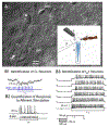
A. Photomicrograph of patched GABAergic negative cell under DIC optics (asterisk). Inset shows configuration for stimulation and recording through a pipette (p). A twisted bipolar stimulating electrode (e) was positioned over incoming afferent fibers in the solitary tract (st). A fiber optic probe was used for optogenetic stimulation (o) and a cannula positioned for drug delivery (c). B1. Cells were categorized as non-GABAergic if they responded with inhibitory post-synaptic potentials to optogenetic stimulation. B2. Action potentials were recorded in response to 20 Hz afferent fiber stimulation. B3. Cells were identified as IA positive (IA+) when the onset of action potentials to a depolarizing stimulus was delayed when preceded by a hyperpolarizing pre-pulse.
Fig. 2.
Fig. 2.
A. Action potentials elicited from the same (IA+) cell after different treatments. Action potentials elicited by afferent stimulation (A1) increased following drug treatment with AmmTX3 (A3). Following optogenetic release of GABA, responses were suppressed both before (A2) and after treatment with AmmTX3 (A4). Resting membrane potentials are shown prior to stimulation for each treatment. B. The mean number of APs elicited by afferent stimulation at 20 Hz for 1 s increased from 6.8 ± 1.3 spikes to 10.4 ± 1.5 spikes (53%) following AmmTX3 in cells with IA (IA+ t-test, P = .001, N = 17) but were unchanged in those without (IAX¯=3.3±0.8 spikes vs X¯=3.4±7.3 spikes; t-test, P = .915, N = 8). A repeated measures Power Test (Systat v. 13) yielded a power value of 0.75, suggesting sufficient power to test the effect of AmmTX3 in IA− cells. Under control conditions (ACSF), the response to afferent stimulation was greater in IA+ cells compared to IA− cells (t-test, P = .028, N: IA+ = 17, IA− = 8). Mean and SEM indicated to the side of data points. The cell illustrated in A is marked with a star.
Fig. 3.
Fig. 3.
A. 4 traces from the same cell showing responses to 150 pA preceded by −60 pA pulse for 450 ms. A delay is evident under control conditions (ACSF) which is suppressed in the presence of AmmTX3. Under dynamic clamp, a delay is again evident, but not when dynamic clamp is turned off. B. Under dynamic clamp, systematic delays were observed in 7 cells: 3 lacking constitutive IA and 4 in which IA was blocked with AmmTX3 (blue line). These delays were comparable to those seen in 10 cells with IA under control (ACSF) conditions (solid red line); ANOVA indicated no difference between delays induced by dynamic clamp and those with constitutive IA (P = .823). In cells without IA, turning dynamic clamp off eliminated the delay (dashed blue line, ANOVA: injected current, P < .001; dynamic clamp, P = .009; current X clamp, P < .001). AmmTX3 eliminated the delay in cells with constitutive IA (dashed red line, ANOVA: injected current, P < .001; AmmTX3, P = .014; current X AmmTX3, P < .001). C. Schematic for implementing dynamic clamp. Membrane voltages (Vm) from cells either lacking constitutive IA or IA blocked with AmmTX3 were used to compute a synthetic IA conductance using IA parameters derived from empirical studies (Chen et al. 2020).
Fig. 4.
Fig. 4.
A. The response of a cell to afferent stimulation under control conditions (dynamic clamp off) was suppressed when a synthetic IA was introduced under dynamic clamp. The response returned to normal when dynamic clamp was turned back off. Traces are continuous in time. B. Effects of a synthetic IA conductance in 11 cells during afferent stimulation at 20 Hz. Six cells lacked constitutive IA (including 2 G+ cells) and 5 had IA blocked pharmacologically with AmmTX3 (including 1 G+ cell). Dynamic clamp parameters were adjusted for G+ and G− cells (see Methods). The effect of dynamic clamp produced a small but consistent suppression to afferent stimulation in 10/11 cells. (dynamic clamp off, X¯=7.0±1.7 spikes/s; dynamic clamp on, X¯=6.1±1.7 spikes/s; t-test: P = .004). Mean and SEM indicated to the side of data points. The cell illustrated in A is indicated by a star in B.
Fig. 5.
Fig. 5.
A. The mean number of APs elicited by afferent stimulation at 20 Hz for 1 s decreased following optogenetic release of GABA for both cells with and without IA (IA+: X¯=6.8±1.3 spikes vs 3.4 ± 1.0 spikes; t-test, P < .001, N = 17; IA−: X¯=3.3±1.5 spikes vs 1.5 ± 0.4 spikes, t-test, P = .034, N = 8). Mean and SEM indicated to the side of data points. Cell illustrated in figure 2A is indicated by a star. B. On average, the response to afferent stimulation was reduced 59% in IA+ cells, whereas responses in cells without IA were suppressed to a nominally smaller degree, 45% (P = .116).
Fig. 6.
Fig. 6.
A. Following optogenetic release of GABA (light) there was a significant reduction in the afferent-evoked response compared to no light (control) in IA+ cells both before and after treatment with AmmTX3. (ANOVA: drug, P <.001; GABA, P < .001, drug × GABA, P = .372, N = 17). B1. Despite the lack of a significant interaction between GABA and AmmTX3 using spike counts, the mean percent suppression due to GABA was smaller following AmmTX3 (paired t-test, P = .025, N = 17). However, this only approached significance when 2 cells with a floor effect (prior to drug treatment) were removed (P = .069, N = 15). B2. Moreover, when the magnitude of inhibition was calculated as the difference in the absolute number of spikes before and after optogenetic stimulation (Δ spikes), AmmTX3 had no effect.
Fig. 7.
Fig. 7.
Photomicrograph of the rostral solitary nucleus stained for P2X2 which effectively demarcates the central subdivision (C). Incoming fibers of the solitary tract (st) were also labeled with P2X2. Many cells with IA were located in the central subdivision (black symbols). Despite the failure to identify cells in the central subdivision without IA in this sample (white symbols), such cells were recorded in our previous study (Chen et al. 2020). An asterisk designates damage to the tissue where a stimulating electrode was positioned adjacent to the solitary tract. Abbreviations: C, central subdivision; IV, 4th ventricle; L, lateral subdivision; M, medial subdivision; V, ventral subdivision.

Similar articles

References

    1. Bailey TW, Hermes SM, Andresen MC, and Aicher SA. Cranial visceral afferent pathways through the nucleus of the solitary tract to caudal ventrolateral medulla or paraventricular hypothalamus: target-specific synaptic reliability and convergence patterns. J Neurosci 26: 11893–11902, 2006. - PMC - PubMed
    1. Bailey TW, Hermes SM, Whittier KL, Aicher SA, and Andresen MC. A-type potassium channels differentially tune afferent pathways from rat solitary tract nucleus to caudal ventrolateral medulla or paraventricular hypothalamus. J Physiol 582: 613–628, 2007. - PMC - PubMed
    1. Bailey TW, Jin YH, Doyle MW, and Andresen MC. Vanilloid-sensitive afferents activate neurons with prominent A-type potassium currents in nucleus tractus solitarius. J Neurosci 22: 8230–8237, 2002. - PMC - PubMed
    1. Bartel DL. Glial responses after chorda tympani nerve injury. J Comp Neurol 520: 2712–2729, 2012. - PMC - PubMed
    1. Beckman ME, and Whitehead MC. Intramedullary connections of the rostral nucleus of the solitary tract in the hamster. Brain Res 557: 265–279, 1991. - PubMed

Publication types

Substances