iBet uBet web content aggregator. Adding the entire web to your favor.


Link to original content: http://pubmed.ncbi.nlm.nih.gov/35254100/
Role of the Transcriptional Regulator ArgR in the Connection between Arginine Metabolism and c-di-GMP Signaling in Pseudomonas putida - PubMed Skip to main page content
U.S. flag

An official website of the United States government

Dot gov

The .gov means it’s official.
Federal government websites often end in .gov or .mil. Before sharing sensitive information, make sure you’re on a federal government site.

Https

The site is secure.
The https:// ensures that you are connecting to the official website and that any information you provide is encrypted and transmitted securely.

Access keys NCBI Homepage MyNCBI Homepage Main Content Main Navigation
. 2022 Apr 12;88(7):e0006422.
doi: 10.1128/aem.00064-22. Epub 2022 Mar 7.

Role of the Transcriptional Regulator ArgR in the Connection between Arginine Metabolism and c-di-GMP Signaling in Pseudomonas putida

Affiliations

Role of the Transcriptional Regulator ArgR in the Connection between Arginine Metabolism and c-di-GMP Signaling in Pseudomonas putida

Laura Barrientos-Moreno et al. Appl Environ Microbiol. .

Abstract

The second messenger cyclic di-GMP (c-di-GMP) is a key molecule that controls different physiological and behavioral processes in many bacteria, including motile-to-sessile lifestyle transitions. Although the external stimuli that modulate cellular c-di-GMP contents are not fully characterized, there is growing evidence that certain amino acids act as environmental cues for c-di-GMP turnover. In the plant-beneficial bacterium Pseudomonas putida KT2440, both arginine biosynthesis and uptake influence second messenger contents and the associated phenotypes. To further understand this connection, we have analyzed the role of ArgR, which in different bacteria is the master transcriptional regulator of arginine metabolism but had not been characterized in P. putida. The results show that ArgR controls arginine biosynthesis and transport, and an argR-null mutant grows poorly with arginine as the sole carbon or nitrogen source and also displays increased biofilm formation and reduced surface motility. Modulation of c-di-GMP levels by exogenous arginine requires ArgR. The expression of certain biofilm matrix components, namely, the adhesin LapF and the exopolysaccharide Pea, as well as the diguanylate cyclase CfcR is influenced by ArgR, likely through the alternative sigma factor RpoS. Our data indicate the existence of a regulatory feedback loop between ArgR and c-di-GMP mediated by FleQ. IMPORTANCE Identifying the molecular mechanisms by which metabolic and environmental signals influence the turnover of the second messenger c-di-GMP is key to understanding the regulation of bacterial lifestyles. The results presented here point at the transcriptional regulator ArgR as a central node linking arginine metabolism and c-di-GMP signaling and indicate the existence of a complex balancing mechanism that connects cellular arginine contents and second messenger levels, ultimately controlling the lifestyles of Pseudomonas putida.

Keywords: Pseudomonas; amino acid biosynthesis; amino acid transport; arginine synthesis; biofilm; c-di-GMP; cell signaling; gene regulation; metabolism; second messenger; transport.


Conflict of interest statement

The authors declare no conflict of interest.

Figures
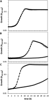
FIG 1
FIG 1
Growth of KT2440 (circles) and the ΔargR mutant (triangles) in M9 minimal medium with 10 mM glucose (A) or 10 mM l-arginine (B) as the carbon source or in M8 medium with 10 mM glucose as the carbon source and 5 mM l-arginine as the nitrogen source (C). Growth was monitored during incubation at 30°C with continuous shaking for 24 h using a Bioscreen apparatus, measuring the absorbance in the 420- to 580-nm range every 30 min. Averages and standard deviations from one representative experiment with three replicates are shown.
FIG 2
FIG 2
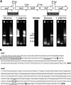
(A) Analysis of the cotranscription of argT and argR with the hisQMP cluster. Electrophoresis was performed on RT-PCR products amplified with primers designed to detect transcripts containing the intergenic regions between argT and hisQ (761 bp) (left) and between hisP and argR (783 bp) (right). Lanes: R, template RNA obtained from P. putida KT2440 cultures; −, negative control without the reverse transcriptase reaction; +, positive control using DNA as the template for the reaction. (B) Determination of the transcription initiation sites for argT and argR. The base corresponding to the +1 site is shaded. The conserved −10 region for the argT promoter and the GC-rich region in the argR promoter are underlined, and the predicted ARG site overlapping the −35 site of argT is boxed. The stop codon for hisP is circled. The square brackets indicate the fragments used to construct transcriptional fusions in plasmids pLBM21 and pLBM20.
FIG 3
FIG 3
Expression of argT::lacZ (pLBM21) (A) and argR::lacZ (pLBM20) (B) during growth in minimal medium with glucose in the absence or presence of l-arginine. β-Galactosidase activity (Miller units) was assessed in KT2440 (wild type [wt]) and the ΔargR mutant harboring pLBM21 or pLBM20 during growth in minimal medium with glucose (light bars) or glucose and 10 mM l-arginine (dark bars). The data correspond to averages and standard deviations from two experiments with three technical replicates each (n = 6). Differences in panel A between the wild type and the mutant and in the wild type in the presence and absence of arginine were statistically significant at all time points, while samples not showing significant differences in panel B at a given time point are indicated as n.s. (not significant) (P ≤ 0.05 by ANOVA).
FIG 4
FIG 4
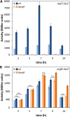
ArgR functions as a repressor of l-arginine biosynthesis genes. β-Galactosidase activities (Miller units) of argG::lacZ (pLBM13) (A) and argH::lacZ (pLBM14) (B) transcriptional fusions were measured in KT2440 (white bars) and the ΔargR mutant (gray bars) in LB at the indicated times. The data are averages and standard deviations from two independent experiments with three technical repetitions each (n = 6). Statistically significant differences between the wild type and the ΔargR mutant were detected from 3 h onward (A) and at 5 h and 7 h (B), respectively (P ≤ 0.05 by Student’s t test).
FIG 5
FIG 5
Influence of ArgR on the changes in cellular c-di-GMP contents in response to exogenous l-arginine. (A) Strain KT2440 (wild type) and the ΔargR mutant harboring pCdrA::gfpC were inoculated into microtiter plates containing diluted LB (1:3) supplied with different final concentrations of l-arginine (0, 5, and 15 mM). Fluorescence and turbidity were recorded every 30 min for 24 h using a Varioskan Lux fluorimeter. Data (GFP counts) correspond to fluorescence values normalized by growth (OD600) and are averages and standard deviations from one representative experiment using three experimental replicates under each condition. The line color intensity indicates increasing concentrations of the amino acid. (B) Values corresponding to the area under the curve derived from fluorescence measurements normalized by culture growth (OD600) were calculated for KT2440 (white bars) and the ΔargR mutant (gray bars), to obtain a global overview of fluorescence along the whole growth curve, where 100% corresponds to the wild type without l-arginine supplementation. Statistically significant differences between groups (P ≤ 0.01 by ANOVA) are indicated by different lowercase letters.
FIG 6
FIG 6
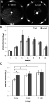
(A) Surface motility of KT2440 and the ΔargR mutant. Cultures grown overnight were diluted to an OD660 of 1, 2 μL was spotted onto the center of the plate, and images were taken after 72 h of growth at 25°C. The assay was done in duplicate, with spotting on six plates in each case, and one representative image is shown for each strain. (B) Biofilm formation by KT2440 (wild type) and its ΔargR derivative during growth in LB in 96-well polystyrene plates under static conditions. At the indicated times, adhered biomass was quantified after crystal violet staining (A595). Data correspond to averages and standard deviations from two independent experiments with 15 replicates per time point. Statistically significant differences between KT2440 and the ΔargR mutant were detected from 5 h onward (P ≤ 0.05 by Student’s t test). (C) Effect of exogenous arginine on biofilm formation by KT2440 (white bars) and the ΔargR mutant (gray bars) after 10 h of growth in FAB medium with glucose as the carbon source supplied with 0, 5, or 15 mM l-arginine. The setup and attached biomass quantification were done as described above for panel B. Data correspond to averages and standard deviations from three independent experiments with three technical replicates. Statistically significant differences (P ≤ 0.05 by ANOVA) are indicated (*).
FIG 7
FIG 7
Influence of argR mutation on the expression of adhesin- and EPS-encoding genes, rpoS and cfcR. Plasmids pMMGA, pMMG1, pMP220-bcs, pMP220-pea, pMP220-peb, and pMIR200, harboring transcriptional fusions of lapA, lapF, pea, peb, bcs, and cfcR to ′lacZ, and pMAMV21, harboring an rpoS-lacZ protein fusion, were introduced into KT2440 (white bars) and the ΔargR mutant (gray bars), and β-galactosidase activity was measured after 10 h of growth in LB. The data are averages and standard deviations from two biological replicates with three technical repetitions each. Statistically significant differences (P ≤ 0.05 by Student’s t test) are indicated (*).
FIG 8
FIG 8
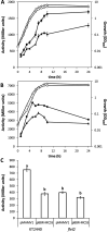
Expression of argR and argT is modulated by c-di-GMP via fleQ. (A and B) Plasmids harboring argR::lacZ (pLBM20) (A) and argT::lacZ (pLBM21) (B) were introduced into KT2440 (circles) and its fleQ mutant derivative cfcK-77 (triangles). Growth (OD600) (open symbols) and β-galactosidase activity (Miller units) (closed symbols) were measured over time in LB. The data are averages and standard deviations from two biological replicates with three technical repetitions each. (C) Activity of the argR::lacZ fusion in KT2440 and cfcK-77 harboring pMAMV1 (cfcR in multicopy) or pBBR1-MCS5 (empty vector) after growth overnight in LB. Cultures were treated with glass beads (diameter of 425 to 600 μm) for 1 min to disrupt cell aggregates formed as a consequence of high c-di-GMP levels due to the presence of pMAMV1. The results correspond to averages and standard deviations from four biological replicates with three technical repetitions each. Statistically significant differences (P ≤ 0.05 by ANOVA) are indicated by different lowercase letters.
FIG 9
FIG 9
Current model of the regulatory network connecting l-arginine metabolism, c-di-GMP signaling, and biofilm formation in P. putida KT2440. ArgR influences c-di-GMP levels (and, therefore, biofilm formation) indirectly through its positive regulatory role in arginine transport and negative effect on arginine synthesis. Arginine and c-di-GMP signaling are connected through the RpoS-dependent transcription of cfcR and potentially via other DGCs. In turn, c-di-GMP modulates the expression of argR via FleQ, thus establishing a feedback loop between arginine pools and c-di-GMP. See the text for further details.

Similar articles

Cited by

References

    1. Römling U, Galperin MY, Gomelsky M. 2013. Cyclic di-GMP: the first 25 years of a universal bacterial second messenger. Microbiol Mol Biol Rev 77:1–52. 10.1128/MMBR.00043-12. - DOI - PMC - PubMed
    1. Chua SL, Tan SY, Rybtke MT, Chen Y, Rice SA, Kjelleberg S, Tolker-Nielsen T, Yang L, Givskov M. 2013. Bis-(3′-5′)-cyclic dimeric GMP regulates antimicrobial peptide resistance in Pseudomonas aeruginosa. Antimicrob Agents Chemother 57:2066–2075. 10.1128/AAC.02499-12. - DOI - PMC - PubMed
    1. Gupta KR, Baloni P, Indi SS, Chatterji D. 2016. Regulation of growth, cell shape, cell division, and gene expression by second messengers (p)ppGpp and cyclic di-GMP in Mycobacterium smegmatis. J Bacteriol 198:1414–1422. 10.1128/JB.00126-16. - DOI - PMC - PubMed
    1. Liang F, Zhang B, Yang Q, Zhang Y, Zheng D, Zhang L, Yan Q, Wu X. 2020. Cyclic-di-GMP regulates the quorum-sensing system and biocontrol activity of Pseudomonas fluorescens 2P24 through the RsmA and RsmE proteins. Appl Environ Microbiol 86:e02016-20. 10.1128/AEM.02016-20. - DOI - PMC - PubMed
    1. Simm R, Morr M, Kader A, Nimtz M, Römling U. 2004. GGDEF and EAL domains inversely regulate cyclic di-GMP levels and transition from sessility to motility. Mol Microbiol 53:1123–1134. 10.1111/j.1365-2958.2004.04206.x. - DOI - PubMed

Publication types

MeSH terms

LinkOut - more resources