iBet uBet web content aggregator. Adding the entire web to your favor.


Link to original content: http://pubmed.ncbi.nlm.nih.gov/35019723/
Rapid SARS-CoV-2 Adaptation to Available Cellular Proteases - PubMed Skip to main page content
U.S. flag

An official website of the United States government

Dot gov

The .gov means it’s official.
Federal government websites often end in .gov or .mil. Before sharing sensitive information, make sure you’re on a federal government site.

Https

The site is secure.
The https:// ensures that you are connecting to the official website and that any information you provide is encrypted and transmitted securely.

Access keys NCBI Homepage MyNCBI Homepage Main Content Main Navigation
. 2022 Mar 9;96(5):e0218621.
doi: 10.1128/jvi.02186-21. Epub 2022 Jan 12.

Rapid SARS-CoV-2 Adaptation to Available Cellular Proteases

Affiliations

Rapid SARS-CoV-2 Adaptation to Available Cellular Proteases

M Zeeshan Chaudhry et al. J Virol. .

Abstract

Recent emergence of SARS-CoV-1 variants demonstrates the potential of this virus for targeted evolution, despite its overall genomic stability. Here we show the dynamics and the mechanisms behind the rapid adaptation of SARS-CoV-2 to growth in Vero E6 cells. The selective advantage for growth in Vero E6 cells is due to increased cleavage efficiency by cathepsins at the mutated S1/S2 site. S1/S2 site also constitutes a heparan sulfate (HS) binding motif that influenced virus growth in Vero E6 cells, but HS antagonist did not inhibit virus adaptation in these cells. The entry of Vero E6-adapted virus into human cells is defective because the mutated spike variants are poorly processed by furin or TMPRSS2. Minor subpopulation that lack the furin cleavage motif in the spike protein rapidly become dominant upon passaging through Vero E6 cells, but wild type sequences are maintained at low percentage in the virus swarm and mediate a rapid reverse adaptation if the virus is passaged again on TMPRSS2+ human cells. Our data show that the spike protein of SARS-CoV-2 can rapidly adapt itself to available proteases and argue for deep sequence surveillance to identify the emergence of novel variants. IMPORTANCE Recently emerging SARS-CoV-2 variants B.1.1.7 (alpha variant), B.1.617.2 (delta variant), and B.1.1.529 (omicron variant) harbor spike mutations and have been linked to increased virus pathogenesis. The emergence of these novel variants highlights coronavirus adaptation and evolution potential, despite the stable consensus genotype of clinical isolates. We show that subdominant variants maintained in the virus population enable the virus to rapidly adapt to selection pressure. Although these adaptations lead to genotype change, the change is not absolute and genomes with original genotype are maintained in the virus swarm. Thus, our results imply that the relative stability of SARS-CoV-2 in numerous independent clinical isolates belies its potential for rapid adaptation to new conditions.

Keywords: SARS-CoV-2; coronavirus spike priming; deep sequencing; furin cleavage site; spike mutation.


Conflict of interest statement

The authors declare no conflict of interest.

Figures
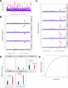
FIG 1
FIG 1
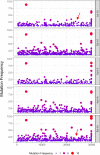
SARS-CoV-2 rapidly adapts upon passage in cultured cells. (A) SARS-CoV-2 with minimal in vitro passaging (Br-P2) was analyzed with deep sequencing and genome diversity is highlighted by plotting genome positions with >1% mutation frequency. (B, C) Serial passages of Braunschweig (Br) or Ischgl (NK) strain were analyzed to assess the composition of viral populations upon passaging in Vero E6 cells. Each symbol represents an individual nucleotide, and genomic positions (x axis) with mutation frequency >1% are plotted. Red arrows highlight the position of the furin cleavage site in the genome. (D) The mutation frequency of nucleotides at the furin cleavage site in the NK strain at indicated passage in Vero E6 cells are plotted on the y axis. Each bar represents one nucleotide for corresponding genomic position from 23,603 to 23,620 (x axis). Dominant mutations are highlighted for certain positions. (E) The sum of mutation frequencies at the furin site position 23,606, 23,607, and 23,616 of NK strain serially passaged on Vero E6 cells is plotted against time. The genomic positions correspond to Wuhan-Hu-1 isolate (GenBank accession no: NC_045512).
FIG 2
FIG 2
Rapid adaption of SARS-CoV-2 is hampered due to absence of subdominant variants at FCS. Plaque purified virus of FI strain was passaged on Vero E6 and virus genomes were analyzed sequentially with deep sequencing to observe genome diversity at each passage. Red arrows highlight the position of the furin cleavage site in the genome. The genomic positions on x axis correspond to Wuhan-Hu-1 isolate (GenBank accession no: NC_045512).
FIG 3
FIG 3
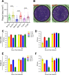
Growth properties of passaged SARS-CoV-2 strains in different cell lines. (A, B) Vero E6 cells were infected with indicated passages of SARS-CoV-2 strains and the plaque size was measured at 72 hpi. Panel A shows the area of virus plaques on Vero E6 cells, and panel B shows representative images of two different wells infected with NK-P4 and NK-P7. (C, D) Virus growth kinetics on Vero E6 cells were established by infecting the cells at an MOI of 0.001. Supernatants (panel C) and cell lysates (panel D) were collected at indicated time points and titrated on Vero E6 cells. (E, F) Calu-3 cells were infected at an MOI of 0.001. Supernatants (panel E) and cell lysates (panel F) were collected at indicated time postinfection and titrated on Vero E6 cells. Data are representative of two independent experiments. Means ±SEM are plotted. Each symbol in panel A represent one plaque and data are pooled from multiple infected wells of two independent experiments. Panel C-F symbols represent biological replicates. Statistical significance was calculated using one way ANOVA with Bonferroni post-test. **P < 0.01; ****P < 0.0001.
FIG 4
FIG 4
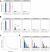
Selection of SARS-CoV-2 subdominant variants with furin cleavage site in TMPRSS2+ human cells. The mutation frequency of genomic positions 23,603 to 23,620 (x axis) in SARS-CoV-2 genome corresponding to furin cleavage site are plotted in panels A, B, and D. Dominant mutation in mutated population are highlighted with arrows. (A) NK strain passaged 6 time in Vero E6 cells (NK6) was serially passaged independently on Calu-3 and Caco-2 cells. P0 vero represents the genome of input virus and P4 calu-3 and P4 caco-2 are the viruses cultured on respective cell lines for four passages. (B) The change in SARS-CoV-2 furin site mutations is shown by plotting the nucleotide mutation frequency of NK6 virus upon serial passaging in Calu-3 cells. (C) The sum of mutation frequency at furin site position 23,606, 23,607, and 23,616 of NK6 strain serially passaged on Calu-3 cells is plotted against time and fitted to the mathematical model. (D) NK strain passaged four times in Vero E6 cells (NK4) was serially passaged on Calu-3 and Caco-2 cells. P0 vero represents the genome of low-passage input virus and P4 calu-3 and P4 caco-2 are the derivative viruses serially passaged on Calu-3 and Caco-2 cell lines for four additional cycles.
FIG 5
FIG 5
Growth inhibition of low and high-passage SARS-CoV-2 by protease inhibitors. Vero E6 (A), Calu-3 (B), and Caco-2 (C) cells were infected at an MOI of 0.01 in the presence of indicated protease inhibitors. The supernatant from infected cells was collected at 24 hpi and titrated on Vero E6 cells. All experiments were twice performed independently with similar results and a typical data set is shown. Three biological replicates per condition are shown as individual dots. Histogram show means and error bars represent ± SEM. Statistical significance was calculated using two-way ANOVA with Dunnett posttest, where untreated control cells served as reference. ns – P > 0.05, *P < 0.05, **P < 0.01, ****P > 0.0001.
FIG 6
FIG 6
Heparan sulfate antagonists fails to inhibit SARS-CoV-2 adaptation on Vero E6 cells. (A) Vero E6 cells were infected with indicated SARS-CoV-2 passaged strains at an MOI of 0.001 and treated with surfen or lactoferrin. The supernatant was collected at 24 hpi and titrated on Vero E6 cells. Data are representative of two independent experiments, and each symbol represents a biological replicate. Mean and ±SEM is plotted. (B) Vero E6 cells were infected with low- or high-passage NK strain viruses. The cells were overlaid with methylcellulose supplemented with 10 μM surfen or 1 mg/mL lactoferrin. The virus plaque size was quantified 72 hpi. Each symbol represents one plaque and data is pooled from multiple infected wells of two independent experiments. (C) NK strain was passaged in Vero E6 cells in the presence of 10 μM surfen. After four passages, virus genome sequence was analyzed with deep sequencing. Each symbol represents an individual nucleotide, and genomic positions (x axis) with mutation frequency >1% are plotted. Red arrow highlights the position of the furin cleavage site and the black arrow shows a dominant silent mutation. Statistical significance was calculated using one way ANOVA test and Bonferroni post-test. ns – P > 0.05; **P < 0.01; ****P > 0.0001.
FIG 7
FIG 7
SARS-CoV-2 mutated spike variants are more efficiently cleaved by cathepsins. (A) Design of the FAM-TAMRA fret pair coupled by the mimetic peptide representing the S1/S2 spike cleavage site. (B) Fluorogenic S1/S2 spike cleavage peptides were cleaved by recombinant proteases in vitro and fold-increase in FAM florescence over time is shown. Data is representative of three independent experiments, and means ± SD of four replicates are plotted. (C) Peptide cleavage efficiency by different recombinant proteases was measured by calculating the area under the curve (AUC) from panel B curves. Technical replicates from three different experiment are pooled and shown as individual dots. Histograms and error bars are means ± SEM. Statistical significance was calculated with one way ANOVA and Bonferroni post-test. ns, P > 0.05; ***P < 0.001; ****P > 0.0001.
FIG 8
FIG 8
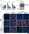
SARS-CoV-2 S furin cleavage site is required for syncytium formation and cell entry into TMPRSS2+ human cells. (A) Vero E6, Calu-3, and Caco-2 cells were infected with pseudotyped VSV harboring VSV-G, wild type, or mutated SARS-CoV-2 spike protein. At 16 hpi, pseudotype entry was analyzed by determining luciferase activity. Signals obtained for particles bearing no envelope protein were used for normalization. The average of three independent experiments is plotted along with ± SEM. (B) Analysis of furin-mediated S protein cleavage. Rhabdoviral particles harboring the indicated S proteins containing a C-terminal HA-tag for detection were lysed and subjected to Western blot analysis. Detection of vesicular stomatitis virus matrix protein (VSV-M) served as control. (C) Syncytium formation assay: A549-ACE2 or A549-ACE2-TMPRSS2 cells were co-transfected with DsRed expressing plasmid and vector that expressed the indicated S proteins (or no S protein, (–) Control). At 24 hpi, cells were incubated in the presence or absence of trypsin (1 μg/mL) for 1 h, before they were fixed, stained with DAPI and analyzed by florescent microscopy (scale bars, 100 μm).

Similar articles

Cited by

References

    1. Wang C, Horby PW, Hayden FG, Gao GF. 2020. A novel coronavirus outbreak of global health concern. Lancet 395:470–473. 10.1016/S0140-6736(20)30185-9. - DOI - PMC - PubMed
    1. Zhu N, Zhang D, Wang W, Li X, Yang B, Song J, Zhao X, Huang B, Shi W, Lu R, Niu P, Zhan F, Ma X, Wang D, Xu W, Wu G, Gao GF, Tan W, China Novel Coronavirus I, Research T . 2020. A novel coronavirus from patients with pneumonia in China, 2019. N Engl J Med 382:727–733. 10.1056/NEJMoa2001017. - DOI - PMC - PubMed
    1. Neuman BW, Adair BD, Yoshioka C, Quispe JD, Orca G, Kuhn P, Milligan RA, Yeager M, Buchmeier MJ. 2006. Supramolecular architecture of severe acute respiratory syndrome coronavirus revealed by electron cryomicroscopy. J Virol 80:7918–7928. 10.1128/JVI.00645-06. - DOI - PMC - PubMed
    1. Tortorici MA, Veesler D. 2019. Structural insights into coronavirus entry. Adv Virus Res 105:93–116. 10.1016/bs.aivir.2019.08.002. - DOI - PMC - PubMed
    1. Hoffmann M, Kleine-Weber H, Schroeder S, Kruger N, Herrler T, Erichsen S, Schiergens TS, Herrler G, Wu NH, Nitsche A, Muller MA, Drosten C, Pohlmann S. 2020. SARS-CoV-2 Cell entry depends on ACE2 and TMPRSS2 and is blocked by a clinically proven protease inhibitor. Cell 181:271–280. 10.1016/j.cell.2020.02.052. - DOI - PMC - PubMed

Publication types

MeSH terms

Supplementary concepts