iBet uBet web content aggregator. Adding the entire web to your favor.


Link to original content: http://pubmed.ncbi.nlm.nih.gov/33564154/
Shaping human telomeres: from shelterin and CST complexes to telomeric chromatin organization - PubMed Skip to main page content
U.S. flag

An official website of the United States government

Dot gov

The .gov means it’s official.
Federal government websites often end in .gov or .mil. Before sharing sensitive information, make sure you’re on a federal government site.

Https

The site is secure.
The https:// ensures that you are connecting to the official website and that any information you provide is encrypted and transmitted securely.

Access keys NCBI Homepage MyNCBI Homepage Main Content Main Navigation
Review
. 2021 Apr;22(4):283-298.
doi: 10.1038/s41580-021-00328-y. Epub 2021 Feb 9.

Shaping human telomeres: from shelterin and CST complexes to telomeric chromatin organization

Affiliations
Review

Shaping human telomeres: from shelterin and CST complexes to telomeric chromatin organization

Ci Ji Lim et al. Nat Rev Mol Cell Biol. 2021 Apr.

Erratum in

Abstract

The regulation of telomere length in mammals is crucial for chromosome end-capping and thus for maintaining genome stability and cellular lifespan. This process requires coordination between telomeric protein complexes and the ribonucleoprotein telomerase, which extends the telomeric DNA. Telomeric proteins modulate telomere architecture, recruit telomerase to accessible telomeres and orchestrate the conversion of the newly synthesized telomeric single-stranded DNA tail into double-stranded DNA. Dysfunctional telomere maintenance leads to telomere shortening, which causes human diseases including bone marrow failure, premature ageing and cancer. Recent studies provide new insights into telomerase-related interactions (the 'telomere replisome') and reveal new challenges for future telomere structural biology endeavours owing to the dynamic nature of telomere architecture and the great number of structures that telomeres form. In this Review, we discuss recently determined structures of the shelterin and CTC1-STN1-TEN1 (CST) complexes, how they may participate in the regulation of telomere replication and chromosome end-capping, and how disease-causing mutations in their encoding genes may affect specific functions. Major outstanding questions in the field include how all of the telomere components assemble relative to each other and how the switching between different telomere structures is achieved.


Conflict of interest statement

Competing interests

T.R.C. is on the board of directors of Merck and a consultant for STORM Therapeutics and Eikon Therapeutics.

Figures

Fig. 1 |
Fig. 1 |. Telomere DNA structures at chromosome ends.
Telomeres are DNA–protein structures at the ends of linear chromosomes. Depicted are the essential telomeric protein complexes shelterin and CTC1–STN1–TEN1 (CST), as well the telomerase RNA–protein complex. The telomeric DNA consists of both double-stranded DNA and single-stranded DNA, comprising in vertebrates the repeat sequence TTAGGG. This duality allows the single-stranded 3′ tail (red) to invade the double-stranded DNA region to form a displacement loop (D-loop) and a telomere loop (T-loop). T-loop formation is regulated by shelterin complexes and can restrict telomerase access to the 3′ tail. Once the telomere is opened up — presumably during the S phase of the cell cycle — telomerase can bind to the 3′ tail through its RNA template (orange) and add telomeric repeats. CST then inhibits telomerase activity, thereby preventing excessive telomere extension. POT1, protection of telomeres protein 1; TIN2, TERF1-interacting nuclear factor 2; TRF1, telomeric repeat-binding factor 1.
Fig. 2 |
Fig. 2 |. Molecular architecture of the human shelterin complex.
a | The human shelterin complex consists of six proteins: telomeric repeat-binding factor 1 (TRF1), TRF2, RAP1, TERF1-interacting nuclear factor 2 (TIN2), TPP1 and protection of telomeres protein 1 (POT1). Shelterin engages both double-stranded DNA (dsDNA) and single-stranded DNA (ssDNA) regions of a telomere. TRF1 and TRF2 are protein homodimers that bind telomeric dsDNA, whereas POT1 binds telomeric ssDNA. b | Map of interactions of shelterin subunits, with binding and structured domains illustrated as boxes. Each polypeptide is displayed from the amino terminus (left) to the carboxy terminus (right) and their stoichiometry is indicated by the number of copies. c | Possible organization of an assembled shelterin complex (excluding RAP1). Unstructured regions are illustrated as coloured lines, depicting the large flexible regions of TRF1 and TRF2 (between their TRF homology (TRFH) domains and Myb domains) and of TPP1 (the C-terminal region after its oligonucleotide/oligosaccharide-binding (OB) domain). The fully assembled shelterin complex comprises two structurally separated modules, which we term the telomere architecture module (TRF1–TIN2–TRF2–RAP1) and the telomerase regulation module (TPP1–POT1). A single TIN2 (TIN2256–276) can interact with the TRFH domain of either TRF1 or TRF2 (dashed arm), whereas the TIN2 N-terminal domain (TIN2N) interacts solely with TRF2. BRCT, BRCA1 carboxy terminal; HJR, Holliday junction resolvase-like; RCT, RAP1 carboxy terminal; TEL patch, TPP1 glutamate (E) and leucine (L)-rich patch.
Fig. 3 |
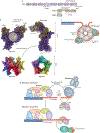
Fig. 3 |. CST and pol α-primase coordination of telomere C-strand fill-in.
a | Interaction map of human CTC1–STN1–TEN1 (CST) complex subunits and their functional sites. b | Atomic structure of CST, derived from cryo-electron microscopy studies (PDB: 6W6W). The magenta halos on the front view indicate two spatially separated groups of residues known to mediate the interactions of CST with the DNA polymerase α-primase (pol α-primase) complex. CST interaction with telomeric single-stranded DNA (ssDNA) can mediate decamerization of CST. c | CST complex decamerization might enable protection of the telomeric G-overhang tail and loading of multiple pol α-primase proteins at a single G-overhang for facilitation of C-strand fill-in. d | Termination of telomerase activity and C-strand fill-in by shelterin, CST and pol α-primase. The shelterin complex recruits telomerase to telomeres and also stimulates extension of the G-overhang by telomerase. Formation of DNA G-quadruplex (G4) structures could prevent telomerase from engaging the tail, but once telomerase has initiated extension, the G4 structure can aid telomerase translocation. The CST complex might prevent telomerase from engaging the G-overhang through competitive binding, or it might disrupt an ongoing extension process by resolving the G4 structure needed for optimal telomerase translocation. Once the extension process is completed, CST can recruit pol α-primase for lagging strand synthesis of the telomeric C-strand to convert the newly synthesized G-overhang into double-stranded DNA. This process is known as telomere C-strand fill-in. OB, oligonucleotide/oligosaccharide binding; STN1c, STN1 carboxy terminal; STN1n, STN1 amino terminal; POT1, protection of telomeres protein 1; TIN2, TERF1-interacting nuclear factor 2; TRF1, telomeric repeat-binding factor 1; wHTH, winged helix–turn–helix motif.
Fig. 4 |
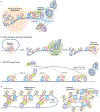
Fig. 4 |. Assembling the telomeres.
a | Telomere maintenance is regulated at two genomic scales: modulation of higher-order DNA–protein architecture across the telomere (gold cloud) and regulation of telomerase activity at the G-overhang (the ‘telomere replisome’; blue cloud). b | The telomeric 3′ tail can be made accessible (green cloud) or reclusive (red clouds) by telomeric protein complexes. The shelterin complex facilitates telomere loop (T-loop) formation, which restricts telomerase access by burying the 3′ tail in double-stranded telomeric DNA. A T-loop can unravel to a linear form to allow telomerase access, which can be further regulated through end-capping by telomeric proteins. c | How TPP1–protection of telomeres protein 1 (POT1) might find the G-overhang among multiple similar binding sites once the tail is made accessible. For simplicity, nucleosomes and other telomeric proteins are omitted from the cartoon. Possible mechanisms of TPP1–POT1 delivery to the G-overhang include 3D diffusion, sliding or hopping along telomeres. Solid and dashed lines indicate stable and transient interactions, respectively. d | Illustrations of two possible and polar opposite outcomes of telomere organization driven by shelterin complexes. A highly ordered shelterin array could result in a zipper-like folding of the telomere. On the other hand, a random deposition of shelterin would result in a disordered telomere architecture. Either possibility restricts access to the telomere 3′ tail and also leads to higher-order telomere organization. CST, CTC1–STN1–TEN1; dsDNA, double-stranded DNA; ssDNA, single-stranded DNA; TIN2, TERF1-interacting nuclear factor 2; TRF1, telomeric repeat-binding factor 1.

Similar articles

Cited by

References

    1. Moyzis RK et al. A highly conserved repetitive DNA sequence, (TTAGGG)n, present at the telomeres of human chromosomes. Proc. Natl Acad. Sci. USA 85, 6622–6626 (1988). - PMC - PubMed
    1. Meyne J, Ratliff RL & Moyzis RK Conservation of the human telomere sequence (TTAGGG)n among vertebrates. Proc. Natl Acad. Sci. USA 86, 7049–7053 (1989). - PMC - PubMed
    1. Makarov VL, Hirose Y & Langmore JP Long G tails at both ends of human chromosomes suggest a C strand degradation mechanism for telomere shortening. Cell 88, 657–666 (1997). - PubMed
    1. Lansdorp PM et al. Heterogeneity in telomere length of human chromosomes. Hum. Mol. Genet 5, 685–691 (1996). - PubMed
    1. Vaziri H et al. Loss of telomeric DNA during aging of normal and trisomy 21 human lymphocytes. Am. J. Hum. Genet 52, 661–667 (1993). - PMC - PubMed

Publication types

LinkOut - more resources