Anatomic and radiologic relationships of neck structures to cervical spine: implications for anterior surgical approaches
- PMID: 33100335
- PMCID: PMC7586192
- DOI: 10.14639/0392-100X-N0503
Anatomic and radiologic relationships of neck structures to cervical spine: implications for anterior surgical approaches
Abstract
Rapporti anatomici e radiologici delle strutture cervicali in relazione al rachide: implicazioni per gli approcci cervicotomici anteriori al rachide.
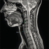
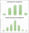
Riassunto: La posizione del framework faringolaringeo è importante nel decidere quale approccio al rachide cervicale adottare. L’obiettivo dello studio è di analizzare la posizione dell’osso ioide e della cartilagine cricoide in relazione ai livelli del rachide cervicale e discuterne le possibili implicazioni per gli accessi cervicotomici al rachide cervicale ed allo spazio prevertebrale. Infatti, per ridurre al minimo le complicanze legate a tale chirurgia ed incrementare l’efficacia del trattamento, la posizione ed estensione del processo patologico a carico del rachide dovrebbe essere inquadrata rispetto ai rapporti anatomici con l’asse faringo-laringeo del singolo paziente. È stata condotta un’analisi retrospettiva di 100 risonanze magnetiche cervicali, escludendo i pazienti che presentavano lesioni che potessero alterare i rapporti anatomici con le vertebre cervicali. Prendendo in esame le scansioni sagittali sulla linea mediana, l’osso ioide ed il margine inferiore della cricoide sono stati proiettati perpendicolarmente sulla superficie anteriore del rachide ed è stata misurata la distanza di queste dal punto di proiezione sulla colonna vertebrale. La distribuzione delle proiezioni dell’osso ioide si attestava tra gli spazi intervertebrali C2-C3 and C4-C5, mentre quella della cartilagine cricoide tra C4-C5 e C7-T1. La distanza media tra queste due strutture era di 49,1 ± 7,7 mm e vi era differenza statisticamente significativa tra sesso maschile e femminile. La posizione della cricoide influenzava significativamente la lunghezza dell’asse faringo-laringeo, mentre la posizione dell’osso ioide era spesso costante. Da quanto osservato, si evince che esista un ampio raggio di variabilità nella posizione dell’osso ioide e della cartilagine cricoidea rispetto ai livelli delle vertebre cervicali. Da ciò ne deriva che una associazione a priori tra specifici livelli cervicali e strutture del collo a rischio sarebbe poco accurata. L’utilizzo dei reperi proposti, facilmente identificabili radiologicamente, può guidare la scelta tra i diversi approcci cervicotomici anteriori al rachide cervicale, riducendo il rischio di complicanze chirurgiche.
Keywords: cervical vertebrae; hypoglossal nerve; recurrent laryngeal nerve; swallowing disorders; vocal cord paralysis.
Plain language summary
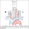
The position of the pharyngolaryngeal framework is very important in choosing the best surgical approach for cervical spine disease. The aim of the present paper is to investigate the position of the hyoid bone and cricoid cartilage in relation to the cervical spine. Moreover, the surgical implications for anterior transcervical approaches to the upper spine and the prevertebral space are discussed. To minimise complication rates and increase surgical effectiveness, the location and extent of the cervical spine disease should be evaluated in the context of the patient’s specific anatomy. A retrospective analysis of 100 cervical spine MRIs was conducted. Patients with diseases that could alter anatomic relationships of cervical structures were excluded. The mid-sagittal view of the hyoid and the inferior margin of the cricoid cartilage were projected perpendicularly to the anterior surface of the cervical vertebrae. The distance between these two landmarks was measured on the same view. The distribution of hyoid projections ranged between C2-C3 and C4-C5 intervertebral space, while the cricoid cartilage ranged between C4-C5 and C7-T1 intervertebral spaces. The mean distance between these two landmarks was 49.1 ± 7.7 mm, with statistically significant differences between males and females. The position of the cricoid cartilage significantly influenced the length of the pharyngolaryngeal framework, while the position of hyoid did not. A wide range of variability in the position of the hyoid bone and the cricoid cartilage in relation to cervical levels exists. This implies that an a priori association of a cervical level to neck structures at risk might be inaccurate. The use of these easily identifiable landmarks on pre-operative imaging may help to guide the choice among different anterior surgical approaches to cervical spine and reduce the risk of surgical complications.
Figures


Similar articles
-
Radiographic Evaluation of the Reliability of Neck Anatomic Structures as Anterior Cervical Surgical Landmarks.World Neurosurg. 2017 Jul;103:133-137. doi: 10.1016/j.wneu.2017.03.129. Epub 2017 Apr 4. World Neurosurg. 2017. PMID: 28385657
-
Reliability and Accuracy of Palpable Anterior Neck Landmarks for the Identification of Cervical Spinal Levels.Asian Spine J. 2018 Feb;12(1):80-84. doi: 10.4184/asj.2018.12.1.80. Epub 2018 Feb 7. Asian Spine J. 2018. PMID: 29503686 Free PMC article.
-
The mandibular angle as a landmark for identification of cervical spinal level.Spine (Phila Pa 1976). 2009 May 1;34(10):1006-11. doi: 10.1097/BRS.0b013e31819f2a03. Spine (Phila Pa 1976). 2009. PMID: 19404175
-
History of Cervical Spine Localization: Surface Landmarks.World Neurosurg. 2024 Nov 2:S1878-8750(24)01824-2. doi: 10.1016/j.wneu.2024.10.119. Online ahead of print. World Neurosurg. 2024. PMID: 39491617 Review.
-
Decision making in the surgical treatment of cervical spine metastases.Spine (Phila Pa 1976). 2009 Oct 15;34(22 Suppl):S108-17. doi: 10.1097/BRS.0b013e3181bae1d2. Spine (Phila Pa 1976). 2009. PMID: 19829270 Review.
References
-
- Bambakidis NC, Dickman CA, Spetzler R, et al. Surgery of the craniovertebral junction. Second Edition New York: Thieme; 2013.
-
- Boriani S, Presutti L, Gasbarrini A, et al. Atlas of craniocervical junction and cervical spine surgery. First Edition New York: Springer; 2017.
-
- Cloward RB. The anterior approach for removal of ruptured cervical disks. J Neurosurg 1958;15:602-17. https://doi.org/10.3171/jns.1958.15.6.0602 10.3171/jns.1958.15.6.0602 - DOI - PubMed
-
- Smith GW, Robinson RA. The treatment of certain cervical-spine disorders by anterior removal of the intervertebral disc and interbody fusion. J Bone Joint Surg Am 1958;40-A:607-24. - PubMed
-
- Southwick WO, Robinson RA. Surgical approaches to the vertebral bodies in the cervical and lumbar regions. J Bone Joint Surg Am 1957;39-A:631-44. - PubMed
MeSH terms
LinkOut - more resources
Full Text Sources
Medical
Miscellaneous


