iBet uBet web content aggregator. Adding the entire web to your favor.


Link to original content: http://pubmed.ncbi.nlm.nih.gov/31636125/
The Chd1 chromatin remodeler forms long-lived complexes with nucleosomes in the presence of ADP·BeF3- and transition state analogs - PubMed Skip to main page content
U.S. flag

An official website of the United States government

Dot gov

The .gov means it’s official.
Federal government websites often end in .gov or .mil. Before sharing sensitive information, make sure you’re on a federal government site.

Https

The site is secure.
The https:// ensures that you are connecting to the official website and that any information you provide is encrypted and transmitted securely.

Access keys NCBI Homepage MyNCBI Homepage Main Content Main Navigation
. 2019 Nov 29;294(48):18181-18191.
doi: 10.1074/jbc.RA119.009782. Epub 2019 Oct 21.

The Chd1 chromatin remodeler forms long-lived complexes with nucleosomes in the presence of ADP·BeF3- and transition state analogs

Affiliations

The Chd1 chromatin remodeler forms long-lived complexes with nucleosomes in the presence of ADP·BeF3- and transition state analogs

Ren Ren et al. J Biol Chem. .

Abstract

Chromatin remodelers use helicase-like ATPase domains to reorganize histone-DNA contacts within the nucleosome. Like other remodelers, the chromodomain helicase DNA-binding protein 1 (Chd1) remodeler repositions nucleosomes by altering DNA topology at its internal binding site on the nucleosome, coupling different degrees of DNA twist and DNA movement to distinct nucleotide-bound states of the ATPase motor. In this work, we used a competition assay to study how variations in the bound nucleotide, Chd1, and the nucleosome substrate affect stability of Chd1-nucleosome complexes. We found that Chd1-nucleosome complexes formed in nucleotide-free or ADP conditions were relatively unstable and dissociated within 30 s, whereas those with the nonhydrolyzable ATP analog AMP-PNP had a mean lifetime of 4.8 ± 0.7 min. Chd1-nucleosome complexes were remarkably stable with ADP·BeF3- and the transition state analogs ADP·AlFX and ADP·MgFX, being resistant to competitor nucleosome over a 24-h period. For the tight ADP·BeF3--stabilized complex, Mg2+ was a critical component that did not freely exchange, and formation of these long-lived complexes had a slow, concentration-dependent step. The ADP·BeF3--stabilized complex did not require the Chd1 DNA-binding domain nor the histone H4 tail and appeared relatively insensitive to sequence differences on either side of the Widom 601 sequence. Interestingly, the complex remained stable in ADP·BeF3- even when nucleosomes contained single-stranded gaps that disrupted most DNA contacts with the guide strand. This finding suggests that binding via the tracking strand alone is sufficient for stabilizing the complex in a hydrolysis-competent state.

Keywords: ATPase; DNA binding protein; chromatin remodeling; chromodomain helicase DNA-binding protein 1 (Chd1); motor protein; nucleoside/nucleotide analog; nucleosome; superfamily 2 (SF2) ATPase; transition state analog; twist defect.


Conflict of interest statement

The authors declare that they have no conflicts of interest with the contents of this article

Figures
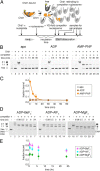
Figure 1.
Figure 1.
A competition assay to determine stability of Chd1–nucleosome complexes. A, schematic workflow of a competition assay to determine off rates for Chd1. B, Chd1–nucleosome complexes are more stable in the presence of the nonhydrolyzable ATP analog AMP-PNP compared with ADP or no nucleotide (apo). As indicated, Chd1 (80 nm) was added to FAM-labeled 12N12 nucleosomes (20 nm), and after a 5-min incubation, reactions were competed with 12N9 unlabeled nucleosomes (2 μm). Lanes 2, 10, and 18 show reactions where unlabeled competitor nucleosome was first mixed with labeled nucleosome prior to addition of Chd1. Binding reactions were resolved on native acrylamide gels. Each gel is a representative of four or more experiments. C, quantification of Chd1–nucleosome dissociation over time. Disappearance of bound complexes in AMP-PNP was fit as a single exponential decay, giving an observed rate of 0.21 ± 0.03 min−1. Stably bound complexes were not detected under apo and ADP conditions. Each point represents the mean from four experiments, with error bars showing S.D. D, competition experiments reveal long-lived Chd1–nucleosome complexes in the presence of transition state analogs ADP·BeF3, ADP·AlFX, and ADP·MgFX. Note that units of time are in hours. For these experiments, Chd1 (40 nm) was added to FAM-labeled 40N40 nucleosomes (10 nm), and after 2-h incubation, the reactions were competed with unlabeled 26N33 nucleosomes (1 μm). Lanes 1, 8, and 15 show reactions where unlabeled competitor nucleosome was first mixed with labeled nucleosome prior to addition of Chd1. Each gel is representative of five or more experiments. E, quantification of Chd1–nucleosome stability over time with transition state analogs. Each point represents an average of five or more experiments, with error bars showing S.D.
Figure 2.
Figure 2.
Effects of buffer components on stability of Chd1–nucleosome transition state complexes. A, excess EDTA does not disrupt long-lived complexes. Competition experiments were performed using 10 nm FAM-labeled 40N40 nucleosomes, 40 nm Chd1, and 1 μm unlabeled 26N33 nucleosomes in the presence of ADP·BeF3. Data points are the mean values ± S.D. from three or more experiments. The “no EDTA” data are equivalent to those shown in Fig. 1D. B, salt and incubation time affect the initial amount of ADP·BeF3–stabilized complex but not its lifetime. For these experiments, 60 nm Chd1 was bound to 20 nm FAM-labeled 12N12 nucleosomes and challenged with 2 μm of unlabeled 12N9 nucleosomes. Each point is the mean ± S.D. from three or more experiments.
Figure 3.
Figure 3.
Time-dependent formation of long-lived Chd1–nucleosome complexes. For these experiments, 40 or 320 nm Chd1 was incubated with 10 nm FAM-labeled 40N40 nucleosomes and challenged with 1 μm unlabeled 26N33 nucleosomes. The apparent on-rates, fit with single exponential equations (dotted traces), were 1.0 ± 0.3 min−1 (320 nm Chd1, 1 min), 0.27 ± 0.08 min−1 (40 nm Chd1, 1 min), and 0.18 ± 0.07 min−1 (40 nm Chd1, 2 h). For the 1-min incubations with competitor, the values are averages of two separate experiments, with error bars indicating the ranges. For the 2-h incubation with competitor, the data are the mean values ± S.D. from four experiments.
Figure 4.
Figure 4.
The Chd1 DNA-binding domain is not required for forming long-lived complexes. A, competition experiments showing stable binding of Chd1(ΔDBD) to nucleosomes in ADP·BeF3. For these experiments, 80 nm Chd1 was incubated with 20 nm FAM-labeled 12N12 nucleosomes and challenged with 2 μm unlabeled 12N9 nucleosomes. Lane 2 shows a reaction where unlabeled competitor was mixed with labeled nucleosome prior to addition of Chd1(ΔDBD). The native acrylamide gel is representative of two experiments at this Chd1(ΔDBD) concentration. B, association of Chd1(ΔDBD) with nucleosomes is time- and concentration-dependent. At three different concentrations of Chd1(ΔDBD), the fraction bound increases over time, with each symbol indicating an average from four experiments, and error bars (often obscured by symbols) indicating S.D. Dotted lines show single exponential fits, with observed rates of 0.09 ± 0.02 min−1 (1280 nm Chd1), 0.021 ± 0.004 min−1 (320 nm Chd1), and 0.005 ± 0.002 min−1 (80 nm Chd1).
Figure 5.
Figure 5.
DNA sequence does not significantly influence formation of long-lived Chd1–nucleosome complexes. A, cartoon schematics of 2:1 Chd1–nucleosome complexes, highlighting the location of the N459C substitution (yellow ovals), which cross-links to nucleosomal DNA. B, cross-linking of Chd1(N459C) to 40N40 nucleosomes in the presence and absence of 12N9 competitor nucleosomes. For each experiment, 300 nm Chd1(N459C) was labeled with azidophencyl bromide, incubated with 150 nm nucleosome for 5 min, and then exposed to 0 or 9 μm 12N9 competitor nucleosome for 1 h before cross-linking. Shown are two experiments performed in 100 mm KCl (lanes 2, 3, 7, and 8) and 150 mm KCl (lanes 4, 5, 9, and 10). The two images show Cy5 (left panel) and FAM (right panel) scans of the same gel. C, quantification of N459C cross-linking observed after addition of competitor nucleosomes. The ratio of peak cross-linking intensities for plus-competitor relative to minus-competitor experiments are plotted as individual points, overlaid on the mean. TA-rich side gives the ratio for Cy5 scans, and TA-poor side gives the ratio for FAM scan. Error bars show S.D. values from eight experiments. n.s., not significant difference.
Figure 6.
Figure 6.
Chd1–nucleosome complexes are relatively stable in ADP·BeF3 despite loss of nucleosomal epitopes. A, molecular rendering of Chd1–nucleosome complex observed by cryoEM (Protein Data Bank code 6FTX) (20), highlighting the ATPase motor (brown and blue), histone H4 tail residues 11–19 (black), and the locations of 9-nt gaps in SHL2.5gapguide (green) and SHL2.5gaptracking (yellow) nucleosomes. In the lower right image, contacts from the ATPase motor to the H4 tail that would be lost are shown as gray surfaces, contacts to DNA that would be lost because of the gaps are shown as yellow or green surfaces, and ATPase contacts to DNA outside the gaps are shown as white surfaces. B, competition experiments in ADP·BeF3 reveal long-lived complexes with nucleosomes containing a ssDNA gap or H4 tail deletion. All FAM-labeled nucleosomes were 12N12 and used at 20 nm, and all competitor nucleosomes were 12N9 added to 2 μm final concentration. For H4Δtail nucleosomes, 80 nm Chd1 was incubated for 5 min prior to competition. For SHL2.5gaptracking nucleosomes, 80 nm Chd1 was incubated for 30 min prior to competition. For SHL2.5gapguide nucleosomes, 200 nm Chd1 was incubated for 30 min prior to competition. Data points are the mean values ± S.D. from three or more experiments. C, an example of a competition experiment with SHL2.5gapguide nucleosomes in ADP·BeF3. Lane 2 shows the background signal when unlabeled competitor nucleosome was mixed with labeled nucleosomes before addition of Chd1. This gel is representative of three experiments, quantified in B. D, competition experiment with SHL2.5gapguide nucleosomes in AMP-PNP. Chd1 (200 nm) was preincubated with 20 nm SHL2.5gapguide nucleosomes for 30 min prior to addition of 2 μm competitor nucleosomes. This gel is representative of four experiments.

Similar articles

Cited by

References

    1. Flaus A., Martin D. M., Barton G. J., and Owen-Hughes T. (2006) Identification of multiple distinct Snf2 subfamilies with conserved structural motifs. Nucleic Acids Res. 34, 2887–2905 10.1093/nar/gkl295 - DOI - PMC - PubMed
    1. Singleton M. R., Dillingham M. S., and Wigley D. B. (2007) Structure and mechanism of helicases and nucleic acid translocases. Annu. Rev. Biochem. 76, 23–50 10.1146/annurev.biochem.76.052305.115300 - DOI - PubMed
    1. Narlikar G. J., Sundaramoorthy R., and Owen-Hughes T. (2013) Mechanisms and functions of ATP-dependent chromatin-remodeling enzymes. Cell 154, 490–503 10.1016/j.cell.2013.07.011 - DOI - PMC - PubMed
    1. Liu F., Putnam A., and Jankowsky E. (2008) ATP hydrolysis is required for DEAD-box protein recycling but not for duplex unwinding. Proc. Natl. Acad. Sci. U.S.A. 105, 20209–20214 10.1073/pnas.0811115106 - DOI - PMC - PubMed
    1. Putnam A. A., and Jankowsky E. (2013) DEAD-box helicases as integrators of RNA, nucleotide and protein binding. Biochim. Biophys. Acta 1829, 884–893 10.1016/j.bbagrm.2013.02.002 - DOI - PMC - PubMed

Publication types

MeSH terms

Associated data