Disentangling isolated dental remains of Asian Pleistocene hominins and pongines
- PMID: 30383758
- PMCID: PMC6211657
- DOI: 10.1371/journal.pone.0204737
Disentangling isolated dental remains of Asian Pleistocene hominins and pongines
Abstract
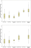
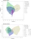
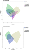
Scholars have debated the taxonomic identity of isolated primate teeth from the Asian Pleistocene for over a century, which is complicated by morphological and metric convergence between orangutan (Pongo) and hominin (Homo) molariform teeth. Like Homo erectus, Pongo once showed considerable dental variation and a wide distribution throughout mainland and insular Asia. In order to clarify the utility of isolated dental remains to document the presence of hominins during Asian prehistory, we examined enamel thickness, enamel-dentine junction shape, and crown development in 33 molars from G. H. R. von Koenigswald's Chinese Apothecary collection (11 Sinanthropus officinalis [= Homo erectus], 21 "Hemanthropus peii," and 1 "Hemanthropus peii" or Pongo) and 7 molars from Sangiran dome (either Homo erectus or Pongo). All fossil teeth were imaged with non-destructive conventional and/or synchrotron micro-computed tomography. These were compared to H. erectus teeth from Zhoukoudian, Sangiran and Trinil, and a large comparative sample of fossil Pongo, recent Pongo, and recent human teeth. We find that Homo and Pongo molars overlap substantially in relative enamel thickness; molar enamel-dentine junction shape is more distinctive, with Pongo showing relatively shorter dentine horns and wider crowns than Homo. Long-period line periodicity values are significantly greater in Pongo than in H. erectus, leading to longer crown formation times in the former. Most of the sample originally assigned to S. officinalis and H. erectus shows greater affinity to Pongo than to the hominin comparative sample. Moreover, enamel thickness, enamel-dentine junction shape, and a long-period line periodicity value in the "Hemanthropus peii" sample are indistinguishable from fossil Pongo. These results underscore the need for additional recovery and study of associated dentitions prior to erecting new taxa from isolated teeth.
Conflict of interest statement
The authors have declared that no competing interests exist.
Figures


Similar articles
-
The early Pleistocene deciduous hominid molar FS-72 from the Sangiran Dome of Java, Indonesia: A taxonomic reappraisal based on its comparative endostructural characterization.Am J Phys Anthropol. 2015 Aug;157(4):666-74. doi: 10.1002/ajpa.22748. Epub 2015 Apr 6. Am J Phys Anthropol. 2015. PMID: 25845703
-
Tooth crown tissue proportions and enamel thickness in Early Pleistocene Homo antecessor molars (Atapuerca, Spain).PLoS One. 2018 Oct 3;13(10):e0203334. doi: 10.1371/journal.pone.0203334. eCollection 2018. PLoS One. 2018. PMID: 30281589 Free PMC article.
-
Variation in enamel thickness within the genus Homo.J Hum Evol. 2012 Mar;62(3):395-411. doi: 10.1016/j.jhevol.2011.12.004. Epub 2012 Feb 22. J Hum Evol. 2012. PMID: 22361504
-
Modern human molar enamel thickness and enamel-dentine junction shape.Arch Oral Biol. 2006 Nov;51(11):974-95. doi: 10.1016/j.archoralbio.2006.04.012. Epub 2006 Jun 30. Arch Oral Biol. 2006. PMID: 16814245 Review.
-
The late Middle Pleistocene hominin fossil record of eastern Asia: synthesis and review.Am J Phys Anthropol. 2010;143 Suppl 51:75-93. doi: 10.1002/ajpa.21442. Am J Phys Anthropol. 2010. PMID: 21086528 Review.
Cited by
-
Oxygen isotopes in orangutan teeth reveal recent and ancient climate variation.Elife. 2024 Mar 8;12:RP90217. doi: 10.7554/eLife.90217. Elife. 2024. PMID: 38457350 Free PMC article.
-
Geometric morphometrics and paleoproteomics enlighten the paleodiversity of Pongo.PLoS One. 2023 Dec 15;18(12):e0291308. doi: 10.1371/journal.pone.0291308. eCollection 2023. PLoS One. 2023. PMID: 38100471 Free PMC article.
-
Measuring Dental Enamel Thickness: Morphological and Functional Relevance of Topographic Mapping.J Imaging. 2023 Jun 23;9(7):127. doi: 10.3390/jimaging9070127. J Imaging. 2023. PMID: 37504804 Free PMC article.
-
Synchrotron X-ray Studies of the Structural and Functional Hierarchies in Mineralised Human Dental Enamel: A State-of-the-Art Review.Dent J (Basel). 2023 Apr 7;11(4):98. doi: 10.3390/dj11040098. Dent J (Basel). 2023. PMID: 37185477 Free PMC article. Review.
-
High-energy synchrotron-radiation-based X-ray micro-tomography enables non-destructive and micro-scale palaeohistological assessment of macro-scale fossil dinosaur bones.J Synchrotron Radiat. 2023 May 1;30(Pt 3):627-633. doi: 10.1107/S1600577523001790. Epub 2023 Apr 7. J Synchrotron Radiat. 2023. PMID: 37026390 Free PMC article.
References
-
- Oakley KP, Campbell BG, Molleson TI, editors. Catalogue of fossil hominids part III: Americas, Asia, Australasia London: British Museum (Natural History); 1975.
-
- Wu X, Poirier FE. Human evolution in China New York: Oxford University Press; 1995.
-
- Antón S. Natural history of Homo erectus. Yearb Phys Anthropol. 2003; 46:126–170. - PubMed
-
- Grine F, Franzen JL. Fossil hominid teeth from the Sangiran Dome (Java, Indonesia). Courier Forschungsinstitut Senckenberg 1994; 171: 75–103.
Publication types
MeSH terms
Grants and funding
LinkOut - more resources
Full Text Sources


