iBet uBet web content aggregator. Adding the entire web to your favor.


Link to original content: http://pubmed.ncbi.nlm.nih.gov/27627203/
Immunodominant SARS Coronavirus Epitopes in Humans Elicited both Enhancing and Neutralizing Effects on Infection in Non-human Primates - PubMed Skip to main page content
U.S. flag

An official website of the United States government

Dot gov

The .gov means it’s official.
Federal government websites often end in .gov or .mil. Before sharing sensitive information, make sure you’re on a federal government site.

Https

The site is secure.
The https:// ensures that you are connecting to the official website and that any information you provide is encrypted and transmitted securely.

Access keys NCBI Homepage MyNCBI Homepage Main Content Main Navigation
. 2016 May 13;2(5):361-76.
doi: 10.1021/acsinfecdis.6b00006. Epub 2016 Apr 11.

Immunodominant SARS Coronavirus Epitopes in Humans Elicited both Enhancing and Neutralizing Effects on Infection in Non-human Primates

Affiliations

Immunodominant SARS Coronavirus Epitopes in Humans Elicited both Enhancing and Neutralizing Effects on Infection in Non-human Primates

Qidi Wang et al. ACS Infect Dis. .

Erratum in

Abstract

Severe acute respiratory syndrome (SARS) is caused by a coronavirus (SARS-CoV) and has the potential to threaten global public health and socioeconomic stability. Evidence of antibody-dependent enhancement (ADE) of SARS-CoV infection in vitro and in non-human primates clouds the prospects for a safe vaccine. Using antibodies from SARS patients, we identified and characterized SARS-CoV B-cell peptide epitopes with disparate functions. In rhesus macaques, the spike glycoprotein peptides S471-503, S604-625, and S1164-1191 elicited antibodies that efficiently prevented infection in non-human primates. In contrast, peptide S597-603 induced antibodies that enhanced infection both in vitro and in non-human primates by using an epitope sequence-dependent (ESD) mechanism. This peptide exhibited a high level of serological reactivity (64%), which resulted from the additive responses of two tandem epitopes (S597-603 and S604-625) and a long-term human B-cell memory response with antisera from convalescent SARS patients. Thus, peptide-based vaccines against SARS-CoV could be engineered to avoid ADE via elimination of the S597-603 epitope. We provide herein an alternative strategy to prepare a safe and effective vaccine for ADE of viral infection by identifying and eliminating epitope sequence-dependent enhancement of viral infection.

Keywords: B-cell peptide epitope; SARS-CoV; antibody-dependent enhancement (ADE); epitope sequence-dependent (ESD) enhancement; peptide; vaccine.


Conflict of interest statement

The authors declare no competing financial interest.

Figures

Figure 1.
Figure 1.. Observation of ADE induced by inactivated SARS-CoV vaccine in the lungs (H&E staining, 200×).
The monkeys were immunized intramuscularly with formalin-inactivated SARS-CoV virions and boosted on day 14 with the same dose of 1.0 mL/monkey (4 × 10⁴ TCID50). The animals were then challenged with nasal cavity inoculation of SARS-CoV (1 × 10⁶ TCID50 in 4.0 mL/monkey, PUMC01 SARS-CoV strain) 14 days after the boost. The animals were sacrificed 6 days after viral challenge, and lung tissues were sampled for general and pathological observations. Pathological examination procedures were as described in reference 37. Infected macaque: Lung interval broadened, visible macrophage infiltration with alveolar epithelial hyperplasia. ADE macaque: Lung interval broadened, lung interval fractured with large amounts of macrophage and lymphocyte infiltration, visible fibrin and protein-rich edema in alveolar cavity. Protected macaques: Lung interval slightly broadened, without visible abnormalities. The arrows indicate the lung lesions of infected and ADE animals.
Figure 2.
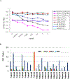
Figure 2.. Immunogenic specificity and long-term memory response of IgG from antisera of SARS patients to S597–625 peptide.
A. The reactivity of human IgG to SARS viral lysates (ELISA kit: Huada S20030004, Beijing, China) is specifically reduced in a dose-dependent manner by S597–625, but not by the hepatitis B virus (HBV) peptide MDIDPYKEFGATVELLSFLP. P223, P73, and P194 represent three antisera from convalescent SARS patients. Reactivity of human IgG with the S597–625 peptide was determined by an ELISA. Lysates (0.025 μg/well) were incubated overnight at room temperature and then blocked by 5% goat serum in PBS (125 μL) for 2 h. 10 μL of antiserum and 100 μL of PBS were further incubated for 30 min at 37°C. Proper amounts of goat anti-human IgG conjugated to HRP were used for detection of OD450 values. Each antiserum was used in duplicate, and the cutoff value was 0.1. B. The antisera of 21 convalescent SARS patients were collected and tested 3 (M3), 12 (M12), 18 (M18), and 24 (M24) months after onset of SARS-CoV infection. The antiserum of patient 9 collected 3 months after onset of infection was omitted due to insufficient sample quantity. The peptides were dissolved in a minimal volume of DMSO and then diluted to a final concentration of 10 μg/mL in carbonate buffer (pH=9.6). In total, 1.0 μg/well was used for capture antibodies from antiserum. The detailed procedure is the same as in Fig. 2A with a cutoff value of 0.1.
Figure 3.
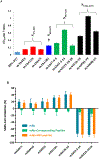
Figure 3.. Generated monoclonal antibodies bound to SARS-CoV virions and shared disparate functions.
A. The defined monoclonal antibodies bound to SARS-CoV virions of SARS-CoV PUMC01 strain (4 × 10 TCID50/mL) . The cutoff value was set to 0.1 (the average for binding of unrelated mAb-HIV-P27). B. Neutralization or enhancement of the SARS-CoV infection in Vero E6 cells by mAbs (1.0 μg/mL) was specifically reduced by the corresponding immunopeptide (0.1 μg/mL), but not by a hepatitis B virus (HBV) peptide (0.1 μg/mL). HBV peptide (LLDYQGMLPV) is an unrelated control peptide from an HBV surface protein. S597–625 and HBV peptide were preincubated with the corresponding mAbs or human antisera for 30 min at 4°C before function was tested. These experiments were performed in triplicate, and the data are presented as the mean ± standard deviation.
Figure 4.
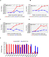
Figure 4.. Antibody-dependent enhancement of SARS-CoV infection.
A, B, C, D: Enhancement of SARS-CoV infection by either mAbs (1.0 μg/mL) or human antisera (1:500) was reduced in a peptide dose-dependent manner. hW49 and hS101 represent two antisera from convalescent SARS patients 3 months after onset. HBV peptide (LLDYQGMLPV) is an unrelated control peptide from HBV surface protein. S597–625 and HBV peptide were preincubated with the corresponding mAb or human antisera for 30 min at 4 °C before function was tested. E. Alanine scanning mutagenesis of the S597–606 showed the minimum requirement (S597–603) for N-terminal binding of S597–625 to mAb43–3-14. This experiment was performed in triplicate, and the data are presented as the mean ± standard deviation.
Figure 5.
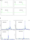
Figure 5.. Tetrameric forms of peptides were successfully synthesized on a branched lysine scaffold.
MAP-S471–503 = [(S471–503)4Lys]2Lys-β-Ala-CONH2; MAP-S604–625 = [(S604–625)4Lys]2Lys-β-Ala-CONH2; MAP-S597–625 = [(S597–625)4Lys]2 Lys-β-Ala-CONH2; MAP-S1164–1191 = [(S1164–1191)4Lys]2Lys-β-Ala-CONH2. RP-HPLC profiles (A) of the MAPs were analyzed by a Shimadzu LC-10AT analytical HPLC system with a C8 column (4.6 mm × 250 mm, 5 μm) and UV detection at 214 nm. The molecular weights (B) were determined by high resolution LC-MS in an Agilent LC/MSD TOF System and hypermass reconstruction of the raw MS data to a single charge.
Figure 6.
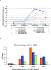
Figure 6.. Rhesus monkeys strongly responded to peptide-based vaccines (Table S5).
Vaccinations (A) were designed as Vac1 (0.9% NaCl) for the control group of four animals, Vac2 (MAPs-S597–625) for the enhancement of SARS-CoV infection in six experimental animals, Vac3 (MAPs-S471–503, MAPs-S604–625 and MAPs-S1164–1191) for the neutralization of SARS-CoV infection in six experimental animals, and Vac4 (MAPs-S471–503, MAPs-S597–625 and MAPs-S1164–1191) for the reduction of neutralizing ability in six experimental animals (Table S4). The average IgG levels against peptides in each vaccine group were monitored by ELISA on the day before injection or boost. IgG level (B) against S604–625 is an average of grouped macaques. The titer of anti-S604–625 IgG was greater than 1:106 in all relevant immunized monkey groups.
Figure 7.
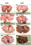
Figure 7.. Peptide-based vaccines neutralized or enhanced SARS-CoV infection of rhesus monkeys.
A. Pathologic changes in lung tissue. The arrows point to the areas that showed pathology. B. Histopathologic examination of macaque lung tissue. Lung damage was pathologically characterized as an average standard grade (table S5). C. Immunohistochemical staining of SARS-CoV-infected cells in lung tissue. Lung tissue sections were incubated with a mixture of mAb4E5, mAb11B1, and mAb9A6 (each at 0.01 μg/mL) and developed using horseradish peroxidase (HRP)-labeled goat anti-mouse IgG (1:1,000 dilution, ZhongShan Inc., Guangzhou, PRC). SARS-CoV-positive cells show brown staining, which localizes in the cytoplasm of monocytes and pneumocytes (magnification 200×). Arrows indicate the lung lesions of animals.
Figure 8.
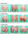
Figure 8.. mAb43-3-14 enhances SARS-CoV infection of rhesus monkeys.
A. Pathologic changes at 6 DPI. B. Histopathologic examination of macaque lung tissues. Lung damage was pathologically characterized as an average standard grade. Control group: grade IV, 0.2 mg/kg group: grade III–IV, and 1.8 mg/kg group: grade IV. C. Immunohistochemical staining of SARS-CoV-infected cells in lung tissue. The staining conditions were the same as in Fig. 6. D. SARS-CoV mRNA in infected monkey lung tissue was quantitatively analyzed from an average of three animals. The data are presented as the geometric mean ± standard deviation. *p < 0.05, **p < 0.01 vs. control group, Δp < 0.05, ΔΔp < 0.01 vs. 0.2 mg/kg group. Arrows indicate the lung lesions of animals.
Scheme 1.
Scheme 1.
Synthesis of multiple antigen peptide systems (MAPS) by chemical ligation of a thiol nucleophilic substitution reaction. B-cell epitopes with a free -SH sequence (containing a Cys residue) were prepared directly. B-cell epitopes without a free -SH group were anchored by an additional Cysteine residue at the C-terminal (Fig S2). A four-branched, brominated, multiple-antigen core peptide (BrK2KA, Fig S3) via lysine two α-amino and side chain amino groups was prepared that finally conjugated with four copies of individual antigenic peptide.

Similar articles

Cited by

References

    1. Hilgenfeld R, Peiris M (2013) From SARS to MERS: 10 years of research on highly pathogenic human coronavirus, Antiviral Res. 100(1): 286–95. - PMC - PubMed
    1. Drosten C, Günther S, Preiser W, van der Werf S, Brodt HR et al. (2003) Identification of a novel coronavirus in patients with severe acute respiratory syndrome. N. Engl. J. Med. 348: 1967–1976. - PubMed
    1. Li W, Shi Z, Yu M, Ren W, Smith C et al. (2003) Bats are natural reservoirs of SARS-like coronaviruses. Science 310: 676–679. - PubMed
    1. Guan Y, Zheng BJ, He YQ, Liu XL, Zhuang ZX, Cheung CL et al. (2003) Isolation and characterization of viruses related to the SARS coronavirus from animals in southern China. Science 302: 276–278. - PubMed
    1. Marra MA, Jones SJ, Astell CR, Holt RA, Brooks-Wilson A et al. (2003) The Genome sequence of the SARS-associated coronavirus. Science 300: 1399–1404. - PubMed

Publication types

MeSH terms