iBet uBet web content aggregator. Adding the entire web to your favor.


Link to original content: http://pubmed.ncbi.nlm.nih.gov/26442282/
Ion-pumping microbial rhodopsins - PubMed Skip to main page content
U.S. flag

An official website of the United States government

Dot gov

The .gov means it’s official.
Federal government websites often end in .gov or .mil. Before sharing sensitive information, make sure you’re on a federal government site.

Https

The site is secure.
The https:// ensures that you are connecting to the official website and that any information you provide is encrypted and transmitted securely.

Access keys NCBI Homepage MyNCBI Homepage Main Content Main Navigation
Review
. 2015 Sep 22:2:52.
doi: 10.3389/fmolb.2015.00052. eCollection 2015.

Ion-pumping microbial rhodopsins

Affiliations
Review

Ion-pumping microbial rhodopsins

Hideki Kandori. Front Mol Biosci. .

Abstract

Rhodopsins are light-sensing proteins used in optogenetics. The word "rhodopsin" originates from the Greek words "rhodo" and "opsis," indicating rose and sight, respectively. Although the classical meaning of rhodopsin is the red-colored pigment in our eyes, the modern meaning of rhodopsin encompasses photoactive proteins containing a retinal chromophore in animals and microbes. Animal and microbial rhodopsins possess 11-cis and all-trans retinal, respectively, to capture light in seven transmembrane α-helices, and photoisomerizations into all-trans and 13-cis forms, respectively, initiate each function. Ion-transporting proteins can be found in microbial rhodopsins, such as light-gated channels and light-driven pumps, which are the main tools in optogenetics. Light-driven pumps, such as archaeal H(+) pump bacteriorhodopsin (BR) and Cl(-) pump halorhodopsin (HR), were discovered in the 1970s, and their mechanism has been extensively studied. On the other hand, different kinds of H(+) and Cl(-) pumps have been found in marine bacteria, such as proteorhodopsin (PR) and Fulvimarina pelagi rhodopsin (FR), respectively. In addition, a light-driven Na(+) pump was found, Krokinobacter eikastus rhodopsin 2 (KR2). These light-driven ion-pumping microbial rhodopsins are classified as DTD, TSA, DTE, NTQ, and NDQ rhodopsins for BR, HR, PR, FR, and KR2, respectively. Recent understanding of ion-pumping microbial rhodopsins is reviewed in this paper.

Keywords: H+ transfer; hydrogen bond; light-driven pump; photocycle; photoisomerizatoin; retinal; structural change.


Figures

Figure 1
Figure 1
Function of rhodopsins. Animal rhodopsins are G-protein coupled receptors, which are categorized as sensors activating a soluble transducer. On the other hand, microbial rhodopsins can act as pumps, channel, and light-sensors. Arrows indicate the direction of transport or flow of a signal. Purple and orange arrows represent energy conversion and signal transduction, respectively.
Figure 2
Figure 2
Chromophore molecules of microbial (left) and animal (right) rhodopsins. β-carotene (top) is the source of the chromophore, and all-trans and 11-cis retinal is bound to protein through the Schiff base linkage.
Figure 3
Figure 3
Phylogenic tree of microbial rhodopsins. This figure is modified from Inoue et al. (2015). The scale bar represents the number of substitutions per site (0.1 indicates 10 nucleotides substitutions per 100 nucleotides). Marine bacterial H+ (yellow), Na+ (orange) and Cl (cyan) pumps have the DTE (or DTX), NDQ, and NTQ motifs, respectively, while archaeal H+ and Cl pumps have the DTD and TSA motifs, respectively. Sensory rhodopsins from halophilic archaea and eubacteria are also shown. AR1, Archaerhodopsin-1; AR2, Archaerhodopsin-2; AR3, Archaerhodopsin-3; HwBR, BR from Haloquadratum walsbyi; MR, Middle rhodopsin; NpHR, HsHR, SrHR, HR from Natronomonas pharaonis; H. salinarum and Salinibacter ruber; HsSRI, HvSRI, SrSRI, sensory rhodopsin I from H. salinarum, Haloarcula vallismortis and S. ruber; HsSRI, NpSRI, sensory rhodopsin I from H. salinarum and N. pharaonis; VsPR, GlPR, NdR1. proteorhodopsins from Vibrio sp. AND4, Gillisia limnaea DSM 15749, Nonlabens dokdonensis DSW-6; XR, xanthorhodopsin, TR, proteorhodopsin from Thermus thermophilus; CbClR, CsClR, SpClR, NmClR, ClR from Citromicrobium bathyomarinum, Citromicrobium sp. JLT1363, Sphingopyxis baekryungensis DSM 16222 and N. marinus; GlNaR, NdNaR, IaNaR, TrNaR1, TrNaR2, NaR from G. limnaea, Nonlabens dokdonensis, Indibacter alkaliphilus, and two NaRs from Truepera radiovictrix, respectively.
Figure 4
Figure 4
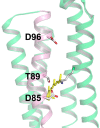
Structure of bacteriorhodopsin (BR) with the DTD motif (PDB: 1QM8, Takeda et al., 1998). The three amino acid residues of the motif (D85, T89, and D96) are located in the C-helix (pink), while other helices are shown in green. Among the seven helices, the A-helix and B-helix are removed to provide a clear view. In BR, D85, and D96 act as the H+ acceptor and donor to the Schiff base, respectively, and T89 forms a hydrogen bond with D85.
Figure 5
Figure 5
Highlighted BR structure with the retinal chromophore, W86, W182, and Y185 (PDB: 1QM8). Y83, W86, and W182 are strongly conserved among the microbial rhodopsins (orange). Aromatic residues are strongly conserved at the Y185, W189, and F219 positions (yellow). In BR, W86, W182, Y185, and W189 constitute the chromophore binding pocket for all-trans retinal (red).
Figure 6
Figure 6
Enlarged structure of BR with the retinal chromophore, W86, W182, and Y185 (PDB: 1QM8). All-trans retinal (A, yellow stick drawing; B, yellow space-filling drawing) is embedded in the binding pocket comprised of these aromatic amino acids.
Figure 7
Figure 7
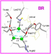
Structure of the Schiff base region in bacteriorhodopsin (BR). This is the side view of the Protein Data Bank structure 1C3W, which has a resolution of 1.55 Å (Luecke et al., 1999). The membrane normal is approximately in the vertical direction of this figure. Hydrogen atoms and hydrogen bonds (dashed lines) are supposed from the structure, while the numbers indicate hydrogen-bonding distances in Å.
Figure 8
Figure 8
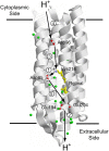
H+ transport pathway in bacteriorhodopsin (BR). Arrows indicate each H+ transfer, and the numbers indicate a temporal order; (1) Schiff base to D85, (2) H+ release, (3) D96 to Schiff base, (4) uptake, and (5) D85 to the H+ release group.
Figure 9
Figure 9
Typical photocycle of microbial rhodopsins showing isomeric and protonation state of retinal. X represents the Schiff base counterion, and D85 in BR also acts as the H+ acceptor from the Schiff base. In a Cl pump such as HR and FR, X is a Cl, so that the M intermediate is not formed because the Schiff base is not deprotonated. Instead, the Cl is transported upwards (in this figure). In KR2, a Na+ pump, X is a D116 acting as the Schiff base counterion and H+ acceptor from the Schiff base. CP and EC indicate cytoplasmic and extracellular domains, respectively. In the unphotolyzed state of microbial rhodopsins, the EC side is generally open through a hydrogen-bonding network but the CP side is closed. While this is persistent in the K and M states, the CP side is open in the N state. When the EC side is closed (black), the CP side is open, as is the case for an ion pump, as occurs in the N intermediate of BR. Such alternative access must work for all H+, Cl, and Na+ pumps.
Figure 10
Figure 10
Schematic drawing of alternative access in BR.
Figure 11
Figure 11
(A) Cl transport pathway in HR and FR (NTQ rhodopsin). Cl binds near the Schiff base region, as is seen from the Cl-dependent color change. The KR2 structure is used in which T is replaced by D116. (B) Na+ transport pathway in KR2 (NDQ rhodopsin). Na+ does not bind near the chromophore in the dark, while a light-induced structural alteration accompanies the uptake of Na+ upon formation of the O intermediate. This is the structure of KR2 (PDB: 3X3C, Kato et al., 2015).
Figure 12
Figure 12
Proposed local structures of KR2 in the dark (A) and in the M intermediate (B), suggested from crystal structures of KR2 at different pHs.

Similar articles

Cited by

References

    1. Agmon N. (1995). The Grotthuss mechanism. Chem. Phys. Lett. 244, 456–462. 10.1016/0009-2614(95)00905-J - DOI
    1. Balashov S. P., Imasheva E. S., Dioumaev A. K., Wang J. M., Jung K. H., Lanyi J. K. (2014). Light-driven Na+ pump from Gillisia limnaea: a high-affinity Na+ binding site is formed transiently in the photocycle. Biochemistry 53, 7549–7561. 10.1021/bi501064n - DOI - PMC - PubMed
    1. Balashov S. P., Petrovskaya L. E., Imasheva E. S., Lukashev E. P., Dioumaev A. K., Wang J. M., et al. . (2013). Breaking the carboxyl rule. Lysine96 facilitates protonation of the Schifff base in the photocycle of a retinal protein from Exiguobacterium sibiricum. J. Biol. Chem. 288, 21254–21265. 10.1074/jbc.M113.465138 - DOI - PMC - PubMed
    1. Béjà O., Aravind L., Koonin E. V., Suzuki M. T., Hadd A., Nguyen L. P., et al. . (2000). Bacterial rhodopsin: evidence for a new type of phototrophy in the sea. Science 289, 1902–1906. 10.1126/science.289.5486.1902 - DOI - PubMed
    1. Béjà O., Lanyi J. K. (2014). Nature's toolkit for microbial rhodopsin ion pumps. Proc. Natl. Acad. Sci. U.S.A. 111, 6538–6539. 10.1073/pnas.1405093111 - DOI - PMC - PubMed