iBet uBet web content aggregator. Adding the entire web to your favor.


Link to original content: http://pubmed.ncbi.nlm.nih.gov/26134649/
Inactivation of Primate Prefrontal Cortex Impairs Auditory and Audiovisual Working Memory - PubMed Skip to main page content
U.S. flag

An official website of the United States government

Dot gov

The .gov means it’s official.
Federal government websites often end in .gov or .mil. Before sharing sensitive information, make sure you’re on a federal government site.

Https

The site is secure.
The https:// ensures that you are connecting to the official website and that any information you provide is encrypted and transmitted securely.

Access keys NCBI Homepage MyNCBI Homepage Main Content Main Navigation
. 2015 Jul 1;35(26):9666-75.
doi: 10.1523/JNEUROSCI.1218-15.2015.

Inactivation of Primate Prefrontal Cortex Impairs Auditory and Audiovisual Working Memory

Affiliations

Inactivation of Primate Prefrontal Cortex Impairs Auditory and Audiovisual Working Memory

Bethany Plakke et al. J Neurosci. .

Abstract

The prefrontal cortex is associated with cognitive functions that include planning, reasoning, decision-making, working memory, and communication. Neurophysiology and neuropsychology studies have established that dorsolateral prefrontal cortex is essential in spatial working memory while the ventral frontal lobe processes language and communication signals. Single-unit recordings in nonhuman primates has shown that ventral prefrontal (VLPFC) neurons integrate face and vocal information and are active during audiovisual working memory. However, whether VLPFC is essential in remembering face and voice information is unknown. We therefore trained nonhuman primates in an audiovisual working memory paradigm using naturalistic face-vocalization movies as memoranda. We inactivated VLPFC, with reversible cortical cooling, and examined performance when faces, vocalizations or both faces and vocalization had to be remembered. We found that VLPFC inactivation impaired subjects' performance in audiovisual and auditory-alone versions of the task. In contrast, VLPFC inactivation did not disrupt visual working memory. Our studies demonstrate the importance of VLPFC in auditory and audiovisual working memory for social stimuli but suggest a different role for VLPFC in unimodal visual processing.

Significance statement: The ventral frontal lobe, or inferior frontal gyrus, plays an important role in audiovisual communication in the human brain. Studies with nonhuman primates have found that neurons within ventral prefrontal cortex (VLPFC) encode both faces and vocalizations and that VLPFC is active when animals need to remember these social stimuli. In the present study, we temporarily inactivated VLPFC by cooling the cortex while nonhuman primates performed a working memory task. This impaired the ability of subjects to remember a face and vocalization pair or just the vocalization alone. Our work highlights the importance of the primate VLPFC in the processing of faces and vocalizations in a manner that is similar to the inferior frontal gyrus in the human brain.

Keywords: monkey; multisensory; visual working memory; vocalization.


Figures

Figure 1.
Figure 1.
Experimental setup. A, The monitor, response button, juice tube, position of the subject, and the bilateral recording cylinders, which were positioned over the PFC, are shown. B, Expanded schematic of the cooling system. The cooling chambers were placed inside the recording cylinders on the subject's head and lie apposed directly to the dural surface overlying VLPFC. Cooled ethanol is pumped through tubing into the cooling chambers. The temperature of the dura is measured with a temperature probe fixed to the bottom of the cooling chamber. Cortical temperature is measured by a different temperature probe inserted through a hole in the center of the cooling chambers into the cortex at a depth of 3 mm. C, Lateral view of the postperfusion brain of Subject 2, with the cylinder location shown by a black circle. Black represents the location of the principal sulcus. Black circle represents the cortical region that was inactivated by cooling on both sides in this subject, as confirmed by presence of dye markers placed into this region before perfusion. D, A coronal MRI section through the region of the VLPFC, which was inactivated in Subject 1. The titanium recording cylinders (which are not visible) cause a shadow and image distortion on the prefrontal cortical surface. Therefore, the region, which was cooled in Subject 1, is approximated by the dotted white outline of the inner edge of the titanium recording cylinders.
Figure 2.
Figure 2.
Schematic of the AV NMTS task. A vocalization movie (with an audio and video track) was presented as the sample stimulus, and the subject was required to remember the auditory and visual components (vocalization and accompanying facial gesture) and to detect the change of either the face or vocalization component in subsequent stimulus presentations with a button press. On Type 1 trials, in half the trials, the nonmatch occurred following the sample and delay period. On Type 2 trials, a matching stimulus intervened and the nonmatch occurred as the third stimulus. In the example shown, the face in the second stimulus of Type 1 trials and the third stimulus of Type 2 trials does not match the sample (visual nonmatch). This occurred for 50% of the trials, and for the other half of trials the audio track was altered and an incongruent vocalization replaced the sample audio track (auditory nonmatch; data not shown).
Figure 3.
Figure 3.
Data from one cooling session showing behavioral accuracy as percentage correct (calculated as cumulative performance, trial by trial, for each block) by block over time. Red line indicates behavioral performance averaging ∼75% correct during the WARM block. Purple line indicates when the cooling process was initiated. Blue line indicates performance when the brain reached a steady temperature and the COLD block began, where accuracy dropped to slightly >50% correct during the COLD block. Each line starts on trial 5 of the block.
Figure 4.
Figure 4.
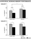
VLPFC inactivation by cooling significantly impairs overall performance accuracy on the AV NMTS task (Experiment 1). *p < 0.05. Mean performance was calculated from 20 testing sessions for each subject with 100 trials (WARM, normal temperature before cooling) and 100 trials during cooling of VLPFC (COLD). Gray bars represent WARM trials. Black bars represent COLD trials (during cooling of the VLPFC). Solid color bars represent performance during auditory component nonmatch trials. Striped bars represent performance during visual component nonmatches. For both subjects, there was a significant decrease in performance during cooling (COLD trials, black bars) compared with the control period (WARM trials, gray bars) for both auditory and visual nonmatch trials. In addition, Subject 1 demonstrated significantly worse performance on auditory trials compared with visual trials. Error bars indicate the SEM calculated across 20 sessions per subject.
Figure 5.
Figure 5.
Percentage accuracy by trial type during AV WM. In both subjects, cooling of the prefrontal cortex resulted in a significant decrease in performance across trial types. A significant interaction between modality and trial type in Subject 1 demonstrated significantly worse performance on auditory Type 1 trials compared with visual trials. *p < 0.05. Colors and shading as in Figure 4. Error bars indicate the SEM calculated across 20 sessions per subject.
Figure 6.
Figure 6.
Inactivation of VLPFC does not significantly affect visual-only WM performance. In Experiment 2 during the visual-only condition, there was no significant change in performance accuracy during cooling of the VLPFC (COLD trials, black bars) compared with the precooling trials (WARM, gray bars) in either subject. Error bars indicate SEM. Mean performance was calculated across 10 sessions (Subject 1) and 11 sessions (Subject 2).
Figure 7.
Figure 7.
Inactivation of VLPFC by cooling significantly decreases auditory-only WM performance. In Experiment 3 during the auditory-only condition, there was a significant decrease in performance for both subjects during the COLD trials (black bars) compared with precooling performance (WARM, gray bars). Error bars indicate SEM. Mean performance was calculated across 10 testing sessions per subject. *p < 0.05.
Figure 8.
Figure 8.
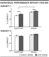
AV performance accuracy without cooling (no temperature change). In Experiment 4, where the temperature was not changed in early and late trial blocks (WARM-WARM), there was no significant difference in performance between the early warm and late warm time periods. Solid color bars represent performance during auditory component nonmatch trials. Striped bars represent performance during visual component nonmatches. For Subject 1, there was an effect of modality indicating better visual performance compared with auditory performance across both time periods. *p < 0.05. White bars represent early warm trials. Gray bars represent late warm trials. Error bars indicate SEM. Mean performance was calculated across 15 sessions per subject.

Similar articles

Cited by

References

    1. Adam R, Noppeney U. Prior auditory information shapes visual category-selectivity in ventral occipito-temporal cortex. Neuroimage. 2010;52:1592–1602. doi: 10.1016/j.neuroimage.2010.05.002. - DOI - PubMed
    1. Bakken HE, Kawasaki H, Oya H, Greenlee JD, Howard MA., 3rd A device for cooling localized regions of human cerebral cortex: technical note. J Neurosurg. 2003;99:604–608. doi: 10.3171/jns.2003.99.3.0604. - DOI - PubMed
    1. Baxter MG, Gaffan D, Kyriazis DA, Mitchell AS. Ventrolateral prefrontal cortex is required for performance of a strategy implementation task but not reinforcer devaluation effects in rhesus monkeys. Eur J Neurosci. 2009;29:2049–2059. doi: 10.1111/j.1460-9568.2009.06740.x. - DOI - PMC - PubMed
    1. Baylis GC, Rolls ET, Leonard CM. Functional subdivisions of the temporal lobe neocortex. J Neurosci. 1987;7:330–342. - PMC - PubMed
    1. Belin P, Zatorre RJ, Lafaille P, Ahad P, Pike B. Voice-selective areas in human auditory cortex. Nature. 2000;403:309–312. doi: 10.1038/35002078. - DOI - PubMed

Publication types

LinkOut - more resources