Effect of Potassium Citrate on Calcium Phosphate Stones in a Model of Hypercalciuria
- PMID: 25855777
- PMCID: PMC4657843
- DOI: 10.1681/ASN.2014121223
Effect of Potassium Citrate on Calcium Phosphate Stones in a Model of Hypercalciuria
Abstract
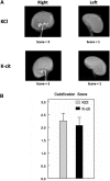
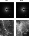
Potassium citrate is prescribed to decrease stone recurrence in patients with calcium nephrolithiasis. Citrate binds intestinal and urine calcium and increases urine pH. Citrate, metabolized to bicarbonate, should decrease calcium excretion by reducing bone resorption and increasing renal calcium reabsorption. However, citrate binding to intestinal calcium may increase absorption and renal excretion of both phosphate and oxalate. Thus, the effect of potassium citrate on urine calcium oxalate and calcium phosphate supersaturation and stone formation is complex and difficult to predict. To study the effects of potassium citrate on urine supersaturation and stone formation, we utilized 95th-generation inbred genetic hypercalciuric stone-forming rats. Rats were fed a fixed amount of a normal calcium (1.2%) diet supplemented with potassium citrate or potassium chloride (each 4 mmol/d) for 18 weeks. Urine was collected at 6, 12, and 18 weeks. At 18 weeks, stone formation was visualized by radiography. Urine citrate, phosphate, oxalate, and pH levels were higher and urine calcium level was lower in rats fed potassium citrate. Furthermore, calcium oxalate and calcium phosphate supersaturation were higher with potassium citrate; however, uric acid supersaturation was lower. Both groups had similar numbers of exclusively calcium phosphate stones. Thus, potassium citrate effectively raises urine citrate levels and lowers urine calcium levels; however, the increases in urine pH, oxalate, and phosphate levels lead to increased calcium oxalate and calcium phosphate supersaturation. Potassium citrate induces complex changes in urine chemistries and resultant supersaturation, which may not be beneficial in preventing calcium phosphate stone formation.
Keywords: hypercalciuria; kidney stones; mineral metabolism.
Copyright © 2015 by the American Society of Nephrology.
Figures


Similar articles
-
Calcium phosphate supersaturation regulates stone formation in genetic hypercalciuric stone-forming rats.Kidney Int. 2000 Feb;57(2):550-60. doi: 10.1046/j.1523-1755.2000.00875.x. Kidney Int. 2000. PMID: 10652032
-
The effect of sodium bicarbonate upon urinary citrate excretion in calcium stone formers.Urology. 2013 Jul;82(1):33-7. doi: 10.1016/j.urology.2013.03.002. Epub 2013 Apr 18. Urology. 2013. PMID: 23602798 Clinical Trial.
-
Dietary treatment of urinary risk factors for renal stone formation. A review of CLU Working Group.Arch Ital Urol Androl. 2015 Jul 7;87(2):105-20. doi: 10.4081/aiua.2015.2.105. Arch Ital Urol Androl. 2015. PMID: 26150027 Review.
-
Short-Term Changes in Urinary Relative Supersaturation Predict Recurrence of Kidney Stones: A Tool to Guide Preventive Measures in Urolithiasis.J Urol. 2018 Nov;200(5):1082-1087. doi: 10.1016/j.juro.2018.06.029. Epub 2018 Jun 22. J Urol. 2018. PMID: 29940247
-
Physiology of acid-base balance: links with kidney stone prevention.Semin Nephrol. 2006 Nov;26(6):441-6. doi: 10.1016/j.semnephrol.2006.10.001. Semin Nephrol. 2006. PMID: 17275581 Review.
Cited by
-
Current Dietary and Medical Prevention of Renal Calcium Oxalate Stones.Int J Gen Med. 2024 Apr 29;17:1635-1649. doi: 10.2147/IJGM.S459155. eCollection 2024. Int J Gen Med. 2024. PMID: 38706742 Free PMC article. Review.
-
Magnesium Decreases Urine Supersaturation but Not Calcium Oxalate Stone Formation in Genetic Hypercalciuric Stone-Forming Rats.Nephron. 2024;148(7):480-486. doi: 10.1159/000534495. Epub 2024 Jan 23. Nephron. 2024. PMID: 38262368
-
Targeting the Liver with Nucleic Acid Therapeutics for the Treatment of Systemic Diseases of Liver Origin.Pharmacol Rev. 2023 Dec 15;76(1):49-89. doi: 10.1124/pharmrev.123.000815. Pharmacol Rev. 2023. PMID: 37696583 Review.
-
Effects of lemon-tomato juice consumption on crystal formation in the urine of patients with calcium oxalate stones: A randomized crossover clinical trial.Curr Urol. 2023 Mar;17(1):25-29. doi: 10.1097/CU9.0000000000000178. Epub 2023 Jan 29. Curr Urol. 2023. PMID: 37692132 Free PMC article.
-
Serum and 24-hour urinary tests cost-effectiveness in stone formers.BMC Urol. 2023 Aug 27;23(1):141. doi: 10.1186/s12894-023-01310-w. BMC Urol. 2023. PMID: 37635222 Free PMC article.
References
-
- Bushinsky DA, Coe FL, Moe OW: Nephrolithiasis. In: The Kidney, edited by Brenner BM, Philadelphia, W.B. Saunders, 2012, pp 1455–1507
-
- Monk RD, Bushinsky DA: Kidney stones. In: Williams Textbook of Endocrinology, edited by Kronenberg HM, Melmed S, Polonsky KS, Larsen PR, Philadelphia, W.B. Saunders, 2011, pp 1350–1367
-
- Monk RD, Bushinsky DA: Nephrolithiasis and nephrocalcinosis. In: Comprehensive Clinical Nephrology, edited by Frehally J, Floege J, Johnson RJ, St. Louis, Elsevier, 2010, pp 687–701
-
- Moe OW, Bonny O: Genetic hypercalciuria. J Am Soc Nephrol 16: 729–745, 2005 - PubMed
-
- Stechman MJ, Loh NY, Thakker RV: Genetics of hypercalciuric nephrolithiasis: Renal stone disease. Ann N Y Acad Sci 1116: 461–484, 2007 - PubMed
Publication types
MeSH terms
Substances
Grants and funding
LinkOut - more resources
Full Text Sources
Other Literature Sources


