iBet uBet web content aggregator. Adding the entire web to your favor.


Link to original content: http://pubmed.ncbi.nlm.nih.gov/25184565/
Microbial peptidyl-prolyl cis/trans isomerases (PPIases): virulence factors and potential alternative drug targets - PubMed Skip to main page content
U.S. flag

An official website of the United States government

Dot gov

The .gov means it’s official.
Federal government websites often end in .gov or .mil. Before sharing sensitive information, make sure you’re on a federal government site.

Https

The site is secure.
The https:// ensures that you are connecting to the official website and that any information you provide is encrypted and transmitted securely.

Access keys NCBI Homepage MyNCBI Homepage Main Content Main Navigation
Review
. 2014 Sep;78(3):544-71.
doi: 10.1128/MMBR.00015-14.

Microbial peptidyl-prolyl cis/trans isomerases (PPIases): virulence factors and potential alternative drug targets

Affiliations
Review

Microbial peptidyl-prolyl cis/trans isomerases (PPIases): virulence factors and potential alternative drug targets

Can M Ünal et al. Microbiol Mol Biol Rev. 2014 Sep.

Abstract

Initially discovered in the context of immunomodulation, peptidyl-prolyl cis/trans isomerases (PPIases) were soon identified as enzymes catalyzing the rate-limiting protein folding step at peptidyl bonds preceding proline residues. Intense searches revealed that PPIases are a superfamily of proteins consisting of three structurally distinguishable families with representatives in every described species of prokaryote and eukaryote and, recently, even in some giant viruses. Despite the clear-cut enzymatic activity and ubiquitous distribution of PPIases, reports on solely PPIase-dependent biological roles remain scarce. Nevertheless, they have been found to be involved in a plethora of biological processes, such as gene expression, signal transduction, protein secretion, development, and tissue regeneration, underscoring their general importance. Hence, it is not surprising that PPIases have also been identified as virulence-associated proteins. The extent of contribution to virulence is highly variable and dependent on the pleiotropic roles of a single PPIase in the respective pathogen. The main objective of this review is to discuss this variety in virulence-related bacterial and protozoan PPIases as well as the involvement of host PPIases in infectious processes. Moreover, a special focus is given to Legionella pneumophila macrophage infectivity potentiator (Mip) and Mip-like PPIases of other pathogens, as the best-characterized virulence-related representatives of this family. Finally, the potential of PPIases as alternative drug targets and first tangible results are highlighted.


Figures

FIG 1
FIG 1
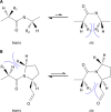
Conformations of peptide bonds. (A) In almost all peptide bonds, the trans conformation is energetically favored because the steric hindrance between the side chains (R1 and R2) of the two consecutive amino acids is the lowest in this conformation. (B) In peptidyl-prolyl bonds, where the α-amino group is part of the side chain, and thus an imide peptide bond is formed, the difference in free energy of the two conformers is comparably smaller. Hence, about 5 to 6% of peptidyl-prolyl bonds are in cis conformation, and the isomerization rate, with half-lives ranging from seconds to minutes, is noticeably long (4, 295).
FIG 2
FIG 2
Representative protein structures for bacterial PPIases. Displayed are structures of the representative members of the three PPIase families of Escherichia coli. (A) PPIase domain of the FKBP FkpA (PDB entry 1Q6U) showing the characteristic α-helix wrapped by a five-stranded β-sheet. The space between the helix and the β-sheet forms the enzymatically active hydrophobic pocket. (B) PPIase domain of the cyclophilin PpiB (PDB entry 2NUL), consisting of the centrally located β-barrel flanked by one α-helix on each side. (C) PPIase domain of the parvulin Par10 (PDB entry 1JNT), organized in the form of a central antiparallel sheet of 4 β-strands with 4 α-helices surrounding it.
FIG 3
FIG 3
Natural FKBP inhibitors and their chemical derivatives. Shown are the structures of FK506 and rapamycin, the two naturally occurring macrolides. Extensive research was done in order to exploit the FKBP binding domain of FK506 (highlighted in yellow) as a lead structure for generating acyclic inhibitors without immunomodulatory side effects. Representatives of each approach with the strongest Ki (app), i.e., 7 nM for SB3 (283), 0.5 nM for VA-10,367 (284), and 2.8 nM for 19d (285), are depicted.
FIG 4
FIG 4
Novel FKBP inhibitors identified by synthetic approaches (A to D) and by screening of databases and compound libraries (E to H), as well as pipecolinic acid derivatives as inhibitors of Mip (I to K). (A) The best alternative to the keto amide group was shown to be a sulfonamide group with a Ki (app) of 160 nM, proving the importance of the keto amide for the protein-ligand interaction (269). (B) In the structurally most divergent PPIase inhibitor, the pipecolinic ring was replaced by a bicyclic ring system which binds to the same hydrophobic cleft, as shown by cocrystallization. Further on, the aromatic ring replaces the pyran, the sulfur atom fills in the pocket usually occupied by the ketone carbonyl group, and the carbonyl group forms a hydrogen bond with Ile56, like the carbonyl of the macrocycle of FK506 does. This compound has a Ki (app) of 7.9 μM (286). (C) A step-by-step approach resulted in the identification of a peptidomimetic inhibitor, β-naphthyl-Pro-Phe-Val-phenyl tetrapeptide, with an IC50 of 0.3 μM (287). (D) Peptidomimetics were generated in a mechanism-based approach. Among the derivatives, a compound resembling a Leu-Pro-tyrosyl tripeptide displayed the highest affinity to FKBP12, with a Ki (app) value of 8.6 μM (288). (E) Using the molecular docking software SANDOCK, N-benzyl-oxycarbonyl-Pro-Pro was identified as the best inhibitor, with a Kd value of 0.8 μM. (F) Steroids such as 5β-pregnan-3,20-dione, with a Kd value of 7 μM, were also detected as novel lead structures for FKBP inhibitors (289). (G) Screening of a library of secondary metabolites resulted in the discovery of cycloheximide as an FKBP12 inhibitor, with an IC50 of 3.6 μM. (H) The cytotoxic effects of cycloheximide were reduced substantially, without remarkably affecting its PPIase-inhibitory property, in the case of the derivative cycloheximide-N-ethylethanoate, with an IC50 of 4.4 μM (290). (I and J) Two lead structures, consisting of a pipecolinic acid ring with either a diketone (I) or a sulfonamide linker (J), were used for further derivatization, and the derivatives were tested in protease-coupled PPIase assays. In accordance with the molecular docking analyses performed in advance, the (S)-enantiomer of structure B showed promising results as an inhibitor, with an IC50 of 6 μM. (K) Replacing the phenyl residue with a benzyl residue led to reduction of the inhibitory capacity (IC50 of 6 μM), underlining the importance of the structural details for a future Mip inhibitor (291).
FIG 5
FIG 5
A close view into the catalytic pocket reveals a high level of structural conservation. Shown is the superimposition of the main amino acid residues building up the catalytically active, hydrophobic pocket of hKBP12 (PDB entry 1FKG; yellow), Mip (PDB entry 1FD9; red), TcMip (PDB entry 1JVW; cyan), and BpML1 (PDB entry 2Y78; green). A remarkable conservation on the amino acid and structural levels can be seen.
FIG 6
FIG 6
Surface topological comparison of Mip and hFKBP12 reveals discriminatory features. Space-filling structural models of hFKBP12 (yellow) (A) and the C-terminal PPIase domain of Legionella Mip (purple) (B) are shown, with views into the respective catalytic pockets. (C) Overlaying both models reveals that the majority of the differences are outside the active site, with a marked difference right in the vicinity of the pocket (highlighted in blue). In hFKBP12, the accessibility of the pocket is sterically hindered, allowing the design of smaller inhibitory molecules with Mip-discriminatory capacity.
FIG 7
FIG 7
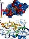
Proposed structural model for the interaction between the PPIase domain of Mip and P290, derived from collagen IV. (A) Average structural model of the Mip77–213-P290 complex derived from NMR measurements. Mip is shown as a blue surface, with residues that were classified as being part of the interaction surface (Γ2 > 12 Hz) colored red. P290 is shown as a stick model. The color coding of P290 reflects the flexibility of the residues according to their calculated B factors, with yellow being rigid and blue being very flexible. (B) Enlarged cutaway view of the Mip binding pocket, where P290 is shown as yellow sticks and Mip is depicted as a blue cartoon, with residues forming the hydrophobic cavity highlighted in green. (Reprinted from reference with permission of the publisher [copyright 2011 Blackwell Publishing Ltd.].)
FIG 8
FIG 8
Structure of D44, a novel inhibitor of plasmodial FKBP35. D44 was identified using structure-based in silico screening of a small-molecule database and verified by in vitro tests.

Similar articles

Cited by

References

    1. Göthel SF, Marahiel MA. 1999. Peptidyl-prolyl cis-trans isomerases, a superfamily of ubiquitous folding catalysts. Cell. Mol. Life Sci. 55:423–436. 10.1007/s000180050299 - DOI - PMC - PubMed
    1. Brandts JF, Halvorson HR, Brennan M. 1975. Consideration of the possibility that the slow step in protein denaturation reactions is due to cis-trans isomerism of proline residues. Biochemistry (Mosc.) 14:4953–4963. 10.1021/bi00693a026 - DOI - PubMed
    1. Cook KH, Schmid FX, Baldwin RL. 1979. Role of proline isomerization in folding of ribonuclease A at low temperatures. Proc. Natl. Acad. Sci. U. S. A. 76:6157–6161. 10.1073/pnas.76.12.6157 - DOI - PMC - PubMed
    1. Stewart DE, Sarkar A, Wampler JE. 1990. Occurrence and role of cis peptide bonds in protein structures. J. Mol. Biol. 214:253–260. 10.1016/0022-2836(90)90159-J - DOI - PubMed
    1. Schmid FX, Baldwin RL. 1978. Acid catalysis of the formation of the slow-folding species of RNase A: evidence that the reaction is proline isomerization. Proc. Natl. Acad. Sci. U. S. A. 75:4764–4768. 10.1073/pnas.75.10.4764 - DOI - PMC - PubMed

Publication types

MeSH terms

LinkOut - more resources