Anatomical transcriptome of G protein-coupled receptors leads to the identification of a novel therapeutic candidate GPR52 for psychiatric disorders
- PMID: 24587241
- PMCID: PMC3938596
- DOI: 10.1371/journal.pone.0090134
Anatomical transcriptome of G protein-coupled receptors leads to the identification of a novel therapeutic candidate GPR52 for psychiatric disorders
Abstract
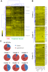
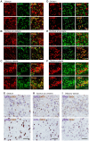
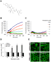
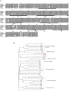
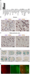
Many drugs of abuse and most neuropharmacological agents regulate G protein-coupled receptors (GPCRs) in the central nervous system (CNS)_ENREF_1. The striatum, in which dopamine D1 and D2 receptors are enriched, is strongly innervated by the ventral tegmental area (VTA), which is the origin of dopaminergic cell bodies of the mesocorticolimbic dopamine system_ENREF_3 and plays a central role in the development of psychiatric disorders_ENREF_4. Here we report the comprehensive and anatomical transcript profiling of 322 non-odorant GPCRs in mouse tissue by quantitative real-time PCR (qPCR), leading to the identification of neurotherapeutic receptors exclusively expressed in the CNS, especially in the striatum. Among them, GPR6, GPR52, and GPR88, known as orphan GPCRs, were shown to co-localize either with a D2 receptor alone or with both D1 and D2 receptors in neurons of the basal ganglia. Intriguingly, we found that GPR52 was well conserved among vertebrates, is Gs-coupled and responsive to the antipsychotic drug, reserpine. We used three types of transgenic (Tg) mice employing a Cre-lox system under the control of the GPR52 promoter, namely, GPR52-LacZ Tg, human GPR52 (hGPR52) Tg, and hGPR52-GFP Tg mice. Detailed histological investigation suggests that GPR52 may modulate dopaminergic and glutamatergic transmission in neuronal circuits responsible for cognitive function and emotion. In support of our prediction, GPR52 knockout and transgenic mice exhibited psychosis-related and antipsychotic-like behaviors, respectively. Therefore, we propose that GPR52 has the potential of being a therapeutic psychiatric receptor. This approach may help identify potential therapeutic targets for CNS diseases.
Conflict of interest statement
Figures


Similar articles
-
Orphan receptor-GPR52 inverse agonist efficacy in ameliorating chronic stress-related deficits in reward motivation and phasic accumbal dopamine activity in mice.Transl Psychiatry. 2024 Sep 7;14(1):363. doi: 10.1038/s41398-024-03081-w. Transl Psychiatry. 2024. PMID: 39242529 Free PMC article.
-
Novel Therapeutic GPCRs for Psychiatric Disorders.Int J Mol Sci. 2015 Jun 19;16(6):14109-21. doi: 10.3390/ijms160614109. Int J Mol Sci. 2015. PMID: 26101869 Free PMC article. Review.
-
Genetic deletion of GPR52 enhances the locomotor-stimulating effect of an adenosine A2A receptor antagonist in mice: A potential role of GPR52 in the function of striatopallidal neurons.Brain Res. 2017 Sep 1;1670:24-31. doi: 10.1016/j.brainres.2017.05.031. Epub 2017 Jun 3. Brain Res. 2017. PMID: 28583861
-
In the Loop: Extrastriatal Regulation of Spiny Projection Neurons by GPR52.ACS Chem Neurosci. 2020 Jul 15;11(14):2066-2076. doi: 10.1021/acschemneuro.0c00197. Epub 2020 Jul 7. ACS Chem Neurosci. 2020. PMID: 32519838
-
Insights into the Promising Prospect of G Protein and GPCR-Mediated Signaling in Neuropathophysiology and Its Therapeutic Regulation.Oxid Med Cell Longev. 2022 Sep 21;2022:8425640. doi: 10.1155/2022/8425640. eCollection 2022. Oxid Med Cell Longev. 2022. PMID: 36187336 Free PMC article. Review.
Cited by
-
Orphan receptor-GPR52 inverse agonist efficacy in ameliorating chronic stress-related deficits in reward motivation and phasic accumbal dopamine activity in mice.Transl Psychiatry. 2024 Sep 7;14(1):363. doi: 10.1038/s41398-024-03081-w. Transl Psychiatry. 2024. PMID: 39242529 Free PMC article.
-
The Orphan G Protein-Coupled Receptor GPR52 is a Novel Regulator of Breast Cancer Multicellular Organization.bioRxiv [Preprint]. 2024 Jul 23:2024.07.22.604482. doi: 10.1101/2024.07.22.604482. bioRxiv. 2024. PMID: 39091857 Free PMC article. Preprint.
-
Exploring orphan GPCRs in neurodegenerative diseases.Front Pharmacol. 2024 Jun 4;15:1394516. doi: 10.3389/fphar.2024.1394516. eCollection 2024. Front Pharmacol. 2024. PMID: 38895631 Free PMC article. Review.
-
Discovery of 3-((4-Benzylpyridin-2-yl)amino)benzamides as Potent GPR52 G Protein-Biased Agonists.J Med Chem. 2024 Jun 13;67(11):9709-9730. doi: 10.1021/acs.jmedchem.4c00856. Epub 2024 May 24. J Med Chem. 2024. PMID: 38788241 Free PMC article.
-
Transcriptomic analysis reveals prolonged neurodegeneration in the hippocampus of adult C57BL/6N mouse deafened by noise.Front Neurosci. 2024 Feb 12;18:1340854. doi: 10.3389/fnins.2024.1340854. eCollection 2024. Front Neurosci. 2024. PMID: 38410162 Free PMC article.
References
-
- Overington JP, Al-Lazikani B, Hopkins AL (2006) How many drug targets are there? Nat Rev Drug Discov 5: 993–996. - PubMed
-
- Gonzalez-Maeso J, Sealfon SC (2009) Agonist-trafficking and hallucinogens. Curr Med Chem 16: 1017–1027. - PubMed
-
- Greengard P (2001) The neurobiology of slow synaptic transmission. Science 294: 1024–1030. - PubMed
-
- Jessell TM, Kandel ER (1993) Synaptic transmission: a bidirectional and self-modifiable form of cell-cell communication. Cell 72 Suppl1–30. - PubMed
-
- Missale C, Nash SR, Robinson SW, Jaber M, Caron MG (1998) Dopamine receptors: from structure to function. Physiol Rev 78: 189–225. - PubMed
MeSH terms
Substances
Grants and funding
LinkOut - more resources
Full Text Sources
Other Literature Sources
Medical
Molecular Biology Databases
Miscellaneous


