A new sauropodomorph dinosaur from the Early Jurassic of Patagonia and the origin and evolution of the sauropod-type sacrum
- PMID: 21298087
- PMCID: PMC3027623
- DOI: 10.1371/journal.pone.0014572
A new sauropodomorph dinosaur from the Early Jurassic of Patagonia and the origin and evolution of the sauropod-type sacrum
Abstract
Background: The origin of sauropod dinosaurs is one of the major landmarks of dinosaur evolution but is still poorly understood. This drastic transformation involved major skeletal modifications, including a shift from the small and gracile condition of primitive sauropodomorphs to the gigantic and quadrupedal condition of sauropods. Recent findings in the Late Triassic-Early Jurassic of Gondwana provide critical evidence to understand the origin and early evolution of sauropods.
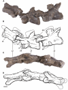
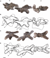
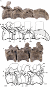
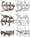
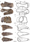
Methodology/principal findings: A new sauropodomorph dinosaur, Leonerasaurus taquetrensis gen. et sp. nov., is described from the Las Leoneras Formation of Central Patagonia (Argentina). The new taxon is diagnosed by the presence of anterior unserrated teeth with a low spoon-shaped crown, amphicoelous and acamerate vertebral centra, four sacral vertebrae, and humeral deltopectoral crest low and medially deflected along its distal half. The phylogenetic analysis depicts Leonerasaurus as one of the closest outgroups of Sauropoda, being the sister taxon of a clade of large bodied taxa composed of Melanorosaurus and Sauropoda.
Conclusions/significance: The dental and postcranial anatomy of Leonerasaurus supports its close affinities with basal sauropods. Despite the small size and plesiomorphic skeletal anatomy of Leonerasaurus, the four vertebrae that compose its sacrum resemble that of the large-bodied primitive sauropods. This shows that the appearance of the sauropod-type of sacrum predated the marked increase in body size that characterizes the origins of sauropods, rejecting a causal explanation and evolutionary linkage between this sacral configuration and body size. Alternative phylogenetic placements of Leonerasaurus as a basal anchisaurian imply a convergent acquisition of the sauropod-type sacrum in the new small-bodied taxon, also rejecting an evolutionary dependence of sacral configuration and body size in sauropodomorphs. This and other recent discoveries are showing that the characteristic sauropod body plan evolved gradually, with a step-wise pattern of character appearance.
Conflict of interest statement
Figures





Similar articles
-
A Giant Dinosaur from the Earliest Jurassic of South Africa and the Transition to Quadrupedality in Early Sauropodomorphs.Curr Biol. 2018 Oct 8;28(19):3143-3151.e7. doi: 10.1016/j.cub.2018.07.063. Epub 2018 Sep 27. Curr Biol. 2018. PMID: 30270189
-
A new basal sauropod dinosaur from the middle Jurassic of Niger and the early evolution of sauropoda.PLoS One. 2009 Sep 16;4(9):e6924. doi: 10.1371/journal.pone.0006924. PLoS One. 2009. PMID: 19756139 Free PMC article.
-
A new basal sauropod from the pre-Toarcian Jurassic of South Africa: evidence of niche-partitioning at the sauropodomorph-sauropod boundary?Sci Rep. 2015 Aug 19;5:13224. doi: 10.1038/srep13224. Sci Rep. 2015. PMID: 26288028 Free PMC article.
-
Biology of the sauropod dinosaurs: the evolution of gigantism.Biol Rev Camb Philos Soc. 2011 Feb;86(1):117-55. doi: 10.1111/j.1469-185X.2010.00137.x. Biol Rev Camb Philos Soc. 2011. PMID: 21251189 Free PMC article. Review.
-
The origin and early evolution of dinosaurs.Biol Rev Camb Philos Soc. 2010 Feb;85(1):55-110. doi: 10.1111/j.1469-185X.2009.00094.x. Epub 2009 Nov 6. Biol Rev Camb Philos Soc. 2010. PMID: 19895605 Review.
Cited by
-
Fertile Goeppertella from the Jurassic of Patagonia: mosaic evolution in the Dipteridaceae-Matoniaceae lineage.AoB Plants. 2023 Jun 7;15(4):plad007. doi: 10.1093/aobpla/plad007. eCollection 2023 Jul. AoB Plants. 2023. PMID: 37426174 Free PMC article.
-
A path to gigantism: Three-dimensional study of the sauropodomorph limb long bone shape variation in the context of the emergence of the sauropod bauplan.J Anat. 2022 Aug;241(2):297-336. doi: 10.1111/joa.13646. Epub 2022 Mar 6. J Anat. 2022. PMID: 35249216 Free PMC article.
-
Walking with early dinosaurs: appendicular myology of the Late Triassic sauropodomorph Thecodontosaurus antiquus.R Soc Open Sci. 2022 Jan 19;9(1):211356. doi: 10.1098/rsos.211356. eCollection 2022 Jan. R Soc Open Sci. 2022. PMID: 35116154 Free PMC article.
-
Extinction of herbivorous dinosaurs linked to Early Jurassic global warming event.Proc Biol Sci. 2020 Nov 25;287(1939):20202310. doi: 10.1098/rspb.2020.2310. Epub 2020 Nov 18. Proc Biol Sci. 2020. PMID: 33203331 Free PMC article.
-
Sacral co-ossification in dinosaurs: The oldest record of fused sacral vertebrae in Dinosauria and the diversity of sacral co-ossification patterns in the group.J Anat. 2021 Apr;238(4):828-844. doi: 10.1111/joa.13356. Epub 2020 Nov 9. J Anat. 2021. PMID: 33164207 Free PMC article.
References
-
- Barrett PM, Upchurch P. Sauropodomorph diversity through time. In: Curry Rogers KA, Wilson JA, editors. The sauropods: evolution and paleobiology. Berkeley: University of California Press; 2005. pp. 125–156.
-
- Wilson JA. Overview of sauropod phylogeny and evolution. In: Curry Rogers KA, Wilson JA, editors. The sauropods: evolution and paleobiology. Berkeley: University of California Press; 2005. pp. 15–49.
-
- Wilson JA, Curry Rogers KA. Introduction: monoliths of the Mesozoic. In: Curry Rogers KA, Wilson JA, editors. The sauropods: evolution and paleobiology. Berkeley: University of California Press; 2005. pp. 1–14.
-
- Colbert EH. Relationships of saurischian dinosaurs. American Museum Novitates. 1964;2181:1–24.
-
- Charig AJ, Attridge J, Crompton AW. On the origin of the sauropods and the classification of the Saurischia. Proceedings of the Linnean Society London. 1965;176:197–221.
Publication types
MeSH terms
LinkOut - more resources
Full Text Sources


