iBet uBet web content aggregator. Adding the entire web to your favor.


Link to original content: http://pubmed.ncbi.nlm.nih.gov/20134403/
ESCRT-II coordinates the assembly of ESCRT-III filaments for cargo sorting and multivesicular body vesicle formation - PubMed Skip to main page content
U.S. flag

An official website of the United States government

Dot gov

The .gov means it’s official.
Federal government websites often end in .gov or .mil. Before sharing sensitive information, make sure you’re on a federal government site.

Https

The site is secure.
The https:// ensures that you are connecting to the official website and that any information you provide is encrypted and transmitted securely.

Access keys NCBI Homepage MyNCBI Homepage Main Content Main Navigation
. 2010 Mar 3;29(5):871-83.
doi: 10.1038/emboj.2009.408. Epub 2010 Feb 4.

ESCRT-II coordinates the assembly of ESCRT-III filaments for cargo sorting and multivesicular body vesicle formation

Affiliations

ESCRT-II coordinates the assembly of ESCRT-III filaments for cargo sorting and multivesicular body vesicle formation

David Teis et al. EMBO J. .

Abstract

The sequential action of five distinct endosomal-sorting complex required for transport (ESCRT) complexes is required for the lysosomal downregulation of cell surface receptors through the multivesicular body (MVB) pathway. On endosomes, the assembly of ESCRT-III is a highly ordered process. We show that the length of ESCRT-III (Snf7) oligomers controls the size of MVB vesicles and addresses how ESCRT-II regulates ESCRT-III assembly. The first step of ESCRT-III assembly is mediated by Vps20, which nucleates Snf7/Vps32 oligomerization, and serves as the link to ESCRT-II. The ESCRT-II subunit Vps25 induces an essential conformational switch that converts inactive monomeric Vps20 into the active nucleator for Snf7 oligomerization. Each ESCRT-II complex contains two Vps25 molecules (arms) that generate a characteristic Y-shaped structure. Mutant 'one-armed' ESCRT-II complexes with a single Vps25 arm are sufficient to nucleate Snf7 oligomerization. However, these oligomers cannot execute ESCRT-III function. Both Vps25 arms provide essential geometry for the assembly of a functional ESCRT-III complex. We propose that ESCRT-II serves as a scaffold that nucleates the assembly of two Snf7 oligomers, which together are required for cargo sequestration and vesicle formation during MVB sorting.


Conflict of interest statement

The authors declare that they have no conflict of interest.

Figures

Figure 1
Figure 1
ESCRT-III regulates the size of MVB vesicles. (A) Transmission electron microscopy of vma4Δ, vma4Δ (THD3)SNF7 and vma4Δ vps25Δ cells. Over-expression of Snf7 (vma4Δ (THD3)SNF7) resulted in the appearance of fewer, larger and more heterogeneous luminal vesicles inside the vacuole of vma4Δ mutant cells. vma4Δ vps25Δ mutants grew very slowly. Representative cells and magnifications are shown. Size bars (500 nm and 100 nm in the magnification). The table shows the range of the number of vesicles detected per vacuole/70 nm section in different ESCRT mutants. (n=30 vacuoles). (B) Average number of MVB vesicles/vacuole/70 nm section (n=30 vacuoles for each sample). vma4Δ mutants have on average 37±15 vesicles, vma4Δ vps25Δ have 0.3±0.5 vesicles and vma4Δ(THD3)SNF7 have 7±3 vesicles/vacuole/section. (C) Distribution of the vesicle diameter in 10 nm step (vma4Δ: n=300, vma4Δ(THD3)SNF7: n=300). In vma4Δ mutants, most vesicle diameters range from 20 to 45 nm (black line). In vma4Δ(THD3)SNF7 mutants, the smallest vesicle is 20 nm and the largest vesicle is 360 nm. In between, the vesicle diameters are evenly distributed.
Figure 2
Figure 2
ESCRT-II is required for Snf7 nucleation. (A) Live cell microscopy of cells harbouring chromosomal integrations for VPS20-GFP and SNF7-mCherry. Size bar (5 μm). Vps20-GFP and Snf7-mCherry co-localize on endosomes. In a vps36Δ mutant Vps20-GFP localizes to endosomes and Snf7-mCherry mis-localized into the cytoplasm. (B) Vps20-GFP localizes to endosomes independently of ESCRT-II (vps36Δ, vps22Δ, vps25Δ triple mutant). Vps20G2A-GFP did not localize to endosomes in WT cells. FM4-64 is shown in red. Size bar (5 μm). (C) Spheroplasts of WT cells harbouring a chromosomal integration of VPS25-Flag or vps36Δ mutants were cross-linked with 5 μm DSP. Solubilized membrane fractions (P13) were subjected to velocity sedimentation and analysed by SDS–PAGE and western blot with the indicated antibodies. *Background band.
Figure 3
Figure 3
Vps20 activation by Vps25 is required for ESCRT-III assembly. (A) Live cell microscopy of vps20Δ, SNF7-GFP cells expressing either VPS20, vps20H1,2, vps20ΔH2 or vps25ΔSNF7-GFP cells expressing vps25T150K. FM4-64 is shown in red. Size bar (5 μm). (B) Spheroplasts of WT or vps20Δ mutants expressing the indicated Vps20 mutants were cross-linked with 5 μM DSP. Solubilized membrane fractions (P13) were subjected to velocity sedimentation and analysed by SDS–PAGE and western blot with the indicated antibodies. (C) Cartoon representation of Chmp3 hVps24 (PDB: 3FRT, (Bajorek et al, 2009)). Indicated are the loop region and helix α1,α2 and auto-inhibitory helix α5, and K61 of Vps20. The auto-inhibitory helix α5 was proposed to move away from the loop region, thereby releasing auto-inhibition. (D) The cytoplasm of cells expressing VPS20-Flag, VPS25-Flag or vps20Δ, vps20loop-HA or vps20Δ, vps25Δ, vps20loop-HA was subjected to size exclusion chromatography on an S200 column. Two fractions were pooled and analysed by SDS–PAGE and western blot with the indicated antibodies. (E) Fluorescence emission spectra of Vps2061−NBD (red trace) and Vps20loop61−NBD (blue trace). (F) In vitro oligomerization experiment. Liposomes were mixed with purified H6-Snf7 alone, or ESCRT-II (E-II), H6-Vps20G2A and H6-Snf7 or ESCRT-II (E-II), H6-Vps20loop and H6-Snf7 (20-fold excess) and incubated for 30 min. The proteins bound to liposomes were solubilized and subjected to velocity sedimentation and analysed by SDS–PAGE and western blot with the indicated antibodies.
Figure 4
Figure 4
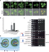
The Vps25–Vps20 nucleation complex controls Snf7 oligomerization during MVB sorting. (A, B) Representative images of cells expressing Mup1-GFP (green). Cells were treated for 60 min with methionine and FM4-64 (red). In snf7Δ vps20Δ or vps20Δ vps20PW cells, Mup1-GFP accumulated on the E-compartment (arrowheads) and was detected at the PM (arrows). In vps24Δ, vps20Δ vps20loop and snf7Δ snx41Δ cells Mup1-GFP was detected only on the class E compartment. Size bar (5 μm). (C) Quantification of the fluorescence ratio (FR) of Mup1-GFP at the PM and on the endosomes in different Vps20 mutants. High FR indicates the accumulation of Mup1-GFP on the E-compartment; n>25 cells for each mutant. (D) Canavanine sensitivity assay with WT cells and the indicated mutants. vps20Δ and snf7Δ were sensitive to 0.6 μg/ml canavanine. vps24Δ, vps2Δ and snf7Δ snx41Δ were more resistant to 0.6 μg/ml canavanine.
Figure 5
Figure 5
A single Vps25 molecule is sufficient activate Vps20. (A) The cartoons depict the Y-shaped structure of ESCRT-II. ESCRT-II consists of one Vps36 (light blue), one Vps22 (dark blue) and two Vps25 molecules/arms (red). The Vps25R83D mutation results in an ‘armless' ESCRT-II complex. Vps36D548R or Vps22D214A mutations generate a ‘one-armed' ESCRT-II complex. Live cell microscopy of Snf7-GFP in the different ESCRT-II mutants. In ESCRT-II mutants, Snf7-GFP no longer localize to the class E compartment. Snf7-GFP eventually aggregated in cytoplasmic dots that are not FM4-64 positive. ‘One armed' ESCRT-II complexes were sufficient for the recruitment of Snf7-GFP to the class E compartment. FM4-64 is shown in red. Size bar (5 μm). (B) Spheroplasts of cells expressing ‘armless' or ‘one-armed' ESCRT-II were cross-linked with 5 μM DSP. Solubilized membrane fractions (P13) were subjected to velocity sedimentation and analysed by SDS–PAGE and western blot with the indicated antibodies.
Figure 6
Figure 6
Two Snf7 filaments are required to trap/encircle cargo. (A) Representative images of cells expressing Mup1-GFP (green). Cells were treated for 60 min with methionine and FM4-64 (red). In all cells, Mup1-GFP accumulates on the E-compartment (arrowheads) and is detected at the PM (arrows). Size bar (5 μm). (B) Fluorescence ratio (FR) of Mup1-GFP at the PM and on the endosomes in different class E mutants; n>25 for each mutant. (C) Canavanine sensitivity assay with WT cells and the indicated mutants. Similar to vps20Δ and snf7Δ, all ESCRT-II mutants (vps36Δ, vps22Δ and vps25Δ) are sensitive to 0.6 μg/ml canavanine. ‘One-armed' and ‘armless' ESCRT-II mutants (Vps22D214A or Vps25R83D) are sensitive to 0.6 μg/ml canavanine. (D) Model for ESCRT-II-mediated assembly of ESCRT-III. Both Vps25 molecules of ESCRT-II would transiently interact with two Vps20 molecules. This interaction induces conformational re-arrangements that simultaneously activate both Vps20 molecules (Step 1). Activated Vps20 then nucleates the oligomerization of two Snf7 filaments (Step 2) that are either branched (I) or run in parallel (II). Capping by Vps24 and Vps2 terminates Snf7 oligomerization (Step 3). Thereby two ESCRT-III filaments could generate an MVB-sorting domain to sequester cargo and form MVB vesicles.

Similar articles

Cited by

References

    1. Alam SL, Langelier C, Whitby FG, Koirala S, Robinson H, Hill CP, Sundquist WI (2006) Structural basis for ubiquitin recognition by the human ESCRT-II EAP45 GLUE domain. Nat Struct Mol Biol 13: 1029–1030 - PubMed
    1. Alam SL, Sun J, Payne M, Welch BD, Blake BK, Davis DR, Meyer HH, Emr SD, Sundquist WI (2004) Ubiquitin interactions of NZF zinc fingers. EMBO J 23: 1411–1421 - PMC - PubMed
    1. Babst M, Katzmann DJ, Estepa-Sabal EJ, Meerloo T, Emr SD (2002a) Escrt-III: an endosome-associated heterooligomeric protein complex required for mvb sorting. Dev Cell 3: 271–282 - PubMed
    1. Babst M, Katzmann DJ, Snyder WB, Wendland B, Emr SD (2002b) Endosome-associated complex, ESCRT-II, recruits transport machinery for protein sorting at the multivesicular body. Dev Cell 3: 283–289 - PubMed
    1. Babst M, Odorizzi G, Estepa EJ, Emr SD (2000) Mammalian tumor susceptibility gene 101 (TSG101) and the yeast homologue, Vps23p, both function in late endosomal trafficking. Traffic 1: 248–258 - PubMed

Publication types

MeSH terms

Substances

LinkOut - more resources