iBet uBet web content aggregator. Adding the entire web to your favor.


Link to original content: http://pubmed.ncbi.nlm.nih.gov/18616971/
Invertebrate muscles: thin and thick filament structure; molecular basis of contraction and its regulation, catch and asynchronous muscle - PubMed Skip to main page content
U.S. flag

An official website of the United States government

Dot gov

The .gov means it’s official.
Federal government websites often end in .gov or .mil. Before sharing sensitive information, make sure you’re on a federal government site.

Https

The site is secure.
The https:// ensures that you are connecting to the official website and that any information you provide is encrypted and transmitted securely.

Access keys NCBI Homepage MyNCBI Homepage Main Content Main Navigation
Review
. 2008 Oct;86(2):72-127.
doi: 10.1016/j.pneurobio.2008.06.004. Epub 2008 Jun 20.

Invertebrate muscles: thin and thick filament structure; molecular basis of contraction and its regulation, catch and asynchronous muscle

Affiliations
Review

Invertebrate muscles: thin and thick filament structure; molecular basis of contraction and its regulation, catch and asynchronous muscle

Scott L Hooper et al. Prog Neurobiol. 2008 Oct.

Abstract

This is the second in a series of canonical reviews on invertebrate muscle. We cover here thin and thick filament structure, the molecular basis of force generation and its regulation, and two special properties of some invertebrate muscle, catch and asynchronous muscle. Invertebrate thin filaments resemble vertebrate thin filaments, although helix structure and tropomyosin arrangement show small differences. Invertebrate thick filaments, alternatively, are very different from vertebrate striated thick filaments and show great variation within invertebrates. Part of this diversity stems from variation in paramyosin content, which is greatly increased in very large diameter invertebrate thick filaments. Other of it arises from relatively small changes in filament backbone structure, which results in filaments with grossly similar myosin head placements (rotating crowns of heads every 14.5 nm) but large changes in detail (distances between heads in azimuthal registration varying from three to thousands of crowns). The lever arm basis of force generation is common to both vertebrates and invertebrates, and in some invertebrates this process is understood on the near atomic level. Invertebrate actomyosin is both thin (tropomyosin:troponin) and thick (primarily via direct Ca(++) binding to myosin) filament regulated, and most invertebrate muscles are dually regulated. These mechanisms are well understood on the molecular level, but the behavioral utility of dual regulation is less so. The phosphorylation state of the thick filament associated giant protein, twitchin, has been recently shown to be the molecular basis of catch. The molecular basis of the stretch activation underlying asynchronous muscle activity, however, remains unresolved.


Figures

Figure 1
Figure 1
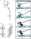
Thin filament structure. A. Schematic showing two actin helices (red, blue), tropomyosin (yellow), and troponin (pink) in vertebrate striated muscle and Lethocerus flight muscle. Modified from Wendt et al. (1997). B. Three dimensional electron microscopy reconstructions of thin filaments interacting with thick filaments at rest (1) and during contraction (2). Panel 3 compares tropomyosin position in panels 1 and 2. Panel 4 shows the likely myosin binding sites. Contour plot, actin; red and yellow helices, tropomyosin; blue dots, myosin binding sites. Modified from Craig and Lehman (2001); data from tarantula.
Figure 2
Figure 2
Myosin, thick filament structure, and actomyosin power stroke. A. Myosin is composed of three paired molecules, the heavy chain and the essential and regulatory light chains. Part of the heavy chains form a coiled coil tail; the remainder of the heavy chains and the two light chains form two globular heads, each of which can independently bind the thin (actin) filament. Modified from Rayment and Holden (1994). B. Thick filaments can be end or side polarized. In end polarized filaments the heads on each half of the filament have the same orientation and the filament thus has a central zone bare of heads. As a result of this orientation, each end of the filament ‘pulls’ the actin filaments with which it interacts toward the central bare zone (arrows). In side polarized filaments the heads on each side of the filament all have the same orientation. Modified from Xu et al. (1996). C. The actomyosin power stroke. 1. A myosin head with bound ADP-Pi approaches an actin binding site. 2. The head become strongly bound. 3. The head rotates about a hinge, and the actin filament is displaced. During this step the Pi disassociates. 4. The ADP also disassociates, ATP binds to the myosin head, and the head dissociates from the actin filament, thus allowing the cycle to repeat. Blue is head catalytic core; yellow and red are, respectively, the pre and post stroke lever arm of the head. Modified from Vale and Milligan (2000).
Figure 3
Figure 3
Explanation of large scale (72 nm) structures often present in large diameter mollusc thick filaments. A. A mollusc thick filament with a checkerboard pattern. B. A reconstituted mollusc paramyosin filament with a simple light-dark banding pattern in which the distance of one repeat unit (one dark and one light band) is 72.5 nm. Schematic shows how an overlap-gap binding of individual paramyosin molecules explains the observed staining pattern (only the gap portions take up the stain). Modified from Cohen (1998).
Figure 4
Figure 4
Conceptual explanation of repeating structures and X-ray analysis. A. A cylinder with objects that are equally spaced around the cylinder in axially repeating groups. Each group has four azimuthally equally spaced objects, each set of objects rotates as one moves axially along the cylinder. Circles linking the objects at each axial level (black circles) and two helices linking nearest objects on different axial levels (blue, red) can be drawn and the distances between them identified. These distances are measured by three of the reflections in an X-ray diffraction pattern. B. The cylinder sliced down the back side and unrolled to form a net. Filled circles are the objects that can be seen in panel A (the objects on the front of the cylinder), open circles are those that cannot be seen (those on the back). The circle at 0 axial distance and 360/0° is grey to indicate that it is a repetition of the object at 0 axial distance and 0/360°.
Figure 5
Figure 5
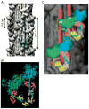
Thick filament strands. A. Surface rendering of three dimensional reconstruction from electron micrographs of tarantula leg muscle. Note helically ascending “hills” (strands) and “valleys”. Blue overlay shows the two myosin heads that form the repeating ‘J’ (dashed line in overlay) motif that forms the strands and a portion of the molecule’s rod in the thick filament body. B. Ribbon representation of two myosin heads (the ‘J’ in panel A) showing that one is free and the other is blocked by binding of its motor domain to the motor domain and essential light chain of the free head. Blue, pink, beige are motor domain, essential light chain, and regulatory light chain of the free head. Green, orange, and yellow are same domains for the blocked head. C. The free head of a crown below binds to the essential light chain of the bound head of the crown above (yellow ellipse). The motor domain of the bound head may also interact with the rod portion of the heads from the crown below (yellow curly bracket). Same color code as in B. Modified, with permission, from Woodhead et al. (2005)
Figure 6
Figure 6
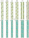
Possible thick filament structures. Top panel (A1–A6). Strands and head origin placements of tarantula leg and Limulus telson (1), lobster abdominal flexor (striated) (2), crab striated (3), lobster smooth (4), scallop adductor (striated) (5), and Lethocerus flight (6) muscle. In each panel small black circles are head origins (where the heads leave the thick filament), yellow helices are strands (composed of interacting heads as shown in Fig. 5), green and red helices connect closest heads on different crowns (red helices in all but A6 hidden by the strands), and arrows indicate heads in angular register. Bottom panel (B1–B6). Possible subfilament organizations consistent with data in A1–A6. Black circles and red and green helices same as in panel A. Black horizontal lines on subfilaments mark 43.5 nm distances measured along the filaments. Every third subfilament colored blue to provide orientation. Scale bar applies to all dimensions (x, y, z) and to both panels.
Figure 7
Figure 7
Thin and thick filament organization and interactions in Lethocerus asynchronous flight muscle. A. Electron micrograph of cross section through muscle (left portion) merging into a schematic of thick (red circles) and thin (blue circles) filaments. Corridors defined by dashed lines mark thin filaments that are in helical register; all thick filaments are also in helical register. Cross bridges are apparent in electron micrograph, inset schematically shows different cross bridge shapes. B. Single thick filament (red circle) and its surrounding thin filaments (small circles). Letters on thick filament indicate head origins, arrow indicates that the filament is right handed. Numbers indicate pairs of thin filaments that are in helical register. Arrow indicates left handed helix of preferred binding sites, helix rotates 60° with each thin filament. C. Spatial relationship between myosin heads (small red ellipses) and thin filament preferred binding sites (large blue ellipses). ‘Unrolled’ and laid flat representation of the arrangement shown in panel B rotated so that the thin filaments (thick vertical blue lines, numbers and blue arrows on top of box) lie in the plane of the figure. Myosin heads leave the thick filament every 14.5 nm, actin helices (leftward slanted thin blue lines) repeat every 38.7 nm (blue arrows, numbers on left of box), preferred thin filament binding sites repeat every 12.9 nm. The thin rightward slanting red lines labeled ‘a–d’ are the right handed helices connecting myosin heads; letters with primes show continuation of helices that have ‘run off’ the right side of the box. D. Average (top panels) and representative individual (bottom panels) three dimensional electron micrograph reconstructions of two thick filaments and an interposed thin filament in rigor (left panels) and pharmacological treatments that reduce thick:thin filament binding (middle and right panels). Numbered red lines on right represent shelf positions, numbered blue lines on left preferred binding sites on the thin filament. In each case lines exactly correspond to those in panel C. Modified from Reedy and Reedy (1985) (A), Wray (1979a) and Schmitz et al. (1994a) (C), and Schmitz et al. (1997) (D).
Figure 8
Figure 8
Explanation of catch. Top four traces: muscle tension; Ca++ concentration and phosphatase activation; cAMP and PKA levels; and twitchin phosphorylation in saline, ACh, saline wash (catch), relaxation induced by 5-HT, ACh and 5-HT, and saline wash. Bottom cartoon, mechanism of catch development and relaxation. Thick filament is bottom thick line, the myosin head is the object resembling a microphone, the thin filament is the row of open circles, and twitchin is the object represented as a coil (unphosphorylated and unable to interact with the thin filament) and straight lines (interacting with the thin filament in alteration with myosin in ACh and continuously during catch in saline). Modified from Funabara et al. (2005) and Butler et al. (2006).

Similar articles

Cited by

References

    1. Abbott BC, Lowy J. Contraction in molluscan smooth muscle. J Physiol. 1958a;141:385–397. - PMC - PubMed
    1. Abbott BC, Lowy J. Mechanical properties of Helix and Mytilus muscle. J Physiol. 1958b;141:398–407. - PMC - PubMed
    1. Abbott RH. An interpretation of the effects of fiber length and calcium on the mechanical properties of insect flight muscle. Cold Spring Harb Symp Quant Biol. 1972;37:647–654.
    1. Abbott RH. The effects of fibre length and calcium ion concentration on the dynamic response of glycerol extracted insect fibrillar muscle. J Physiol. 1973;231:195–208. - PMC - PubMed
    1. Abbott RH, Cage PE. A possible mechanism of length activation in insect fibrillar flight muscle. J Muscle Res Cell Motil. 1984;5:387–397. - PubMed

Publication types