The lumbar spine in Neanderthals shows natural kyphosis
- PMID: 18301930
- PMCID: PMC2525911
- DOI: 10.1007/s00586-008-0640-y
The lumbar spine in Neanderthals shows natural kyphosis
Abstract
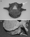
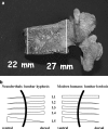
Nowadays, lumbar spondylosis is one of the most frequent causes of lower back pain. In order to improve our understanding of the lumbar spine anatomy and functionality over time, we compared the lumbar vertebrae of Neanderthals with those of anatomically modern humans. The fossil record reports on only two Neanderthal skeletons (i.e., Kebara 2 and Shanidar 3, both predating the appearance of modern humans) with full preservation of the entire lumbar spine. Examination of these early hominids showed that they display natural lumbar kyphosis, with only mild degenerative changes of the lumbar spine (ages at death: 30-35 years, Kebara 2; and 35-50 years, Shanidar 3). This finding is highly unexpected since Neanderthals are known to have had extraordinary physical activity due to demanding living conditions. The adult lumbar spines discussed here therefore show no correlation between high physical activity and degenerative spine disease as known from recent times. We speculate that both the kyphosis itself and the massive and heavily muscled skeleton of Neanderthals are causative for the minimal bone degeneration. We conclude that a kyphotic lumbar spine is the natural anatomy in these two Neanderthal individuals. Future research will reveal if this holds true for the entire Neanderthal species.
Figures


Similar articles
-
Morphology and function of the lumbar spine of the Kebara 2 Neandertal.Am J Phys Anthropol. 2010 Aug;142(4):549-57. doi: 10.1002/ajpa.21256. Am J Phys Anthropol. 2010. PMID: 20091808
-
How do anterior/posterior translations of the thoracic cage affect the sagittal lumbar spine, pelvic tilt, and thoracic kyphosis?Eur Spine J. 2002 Jun;11(3):287-93. doi: 10.1007/s00586-001-0350-1. Epub 2001 Nov 1. Eur Spine J. 2002. PMID: 12107799 Free PMC article.
-
Midlumbar lateral flexion stability measured in healthy volunteers by in vivo fluoroscopy.Spine (Phila Pa 1976). 2009 Oct 15;34(22):E811-7. doi: 10.1097/BRS.0b013e3181b1feba. Spine (Phila Pa 1976). 2009. PMID: 19829245
-
Spinopelvic pathways to bipedality: why no hominids ever relied on a bent-hip-bent-knee gait.Philos Trans R Soc Lond B Biol Sci. 2010 Oct 27;365(1556):3289-99. doi: 10.1098/rstb.2010.0112. Philos Trans R Soc Lond B Biol Sci. 2010. PMID: 20855303 Free PMC article. Review.
-
Between a rock and a cold place: Neanderthal biocultural cold adaptations.Evol Anthropol. 2021 Jul;30(4):262-279. doi: 10.1002/evan.21894. Epub 2021 Apr 2. Evol Anthropol. 2021. PMID: 33797824 Review.
Cited by
-
Inferring lumbar lordosis in Neandertals and other hominins.PNAS Nexus. 2022 Mar 2;1(1):pgab005. doi: 10.1093/pnasnexus/pgab005. eCollection 2022 Mar. PNAS Nexus. 2022. PMID: 36712807 Free PMC article.
-
Predicted Archaic 3D Genome Organization Reveals Genes Related to Head and Spinal Cord Separating Modern from Archaic Humans.Cells. 2019 Dec 24;9(1):48. doi: 10.3390/cells9010048. Cells. 2019. PMID: 31878147 Free PMC article.
-
Morphology, pathology, and the vertebral posture of the La Chapelle-aux-Saints Neandertal.Proc Natl Acad Sci U S A. 2019 Mar 12;116(11):4923-4927. doi: 10.1073/pnas.1820745116. Epub 2019 Feb 25. Proc Natl Acad Sci U S A. 2019. PMID: 30804177 Free PMC article.
-
Clinical Decision-Making in Chronic Spine Pain: Dilemma of Image-Based Diagnosis of Degenerative Spine and Generation Mechanisms for Nociceptive, Radicular, and Referred Pain.Biomed Res Int. 2018 Dec 17;2018:8793843. doi: 10.1155/2018/8793843. eCollection 2018. Biomed Res Int. 2018. PMID: 30648110 Free PMC article.
-
Longitudinal study of radiographic spinal osteoarthritis in a macaque model.J Orthop Res. 2011 Aug;29(8):1152-60. doi: 10.1002/jor.21390. Epub 2011 Mar 4. J Orthop Res. 2011. PMID: 21381096 Free PMC article.
References
-
- Aeby C. Die Altersverschiedenheiten der menschlichen Wirbelsäule. Arch Anat Physiol. 1879;10:77.
-
- Arensburg B, Bar Yosef O, Chech M, Goldberg P, Laville H, Meignen L, Rak Y, Tchernov E, Tillier AM, Vandermeersch B Une sépulture néanderthalien dans la grotte de Kebara (Israel) (1985) Compte Rendus des Séances de l´Académie des Science (Paris), Série II 300:227–230
-
- Arensburg B (1991) The vertebral column, thoracic cage and hyoid bone. In: Bar Yosef O. and Vandermeersch B. Le Squelette Mousterian de Kebara 2:113–146. Editions du C.N.R.S. (Paris)
Publication types
MeSH terms
LinkOut - more resources
Full Text Sources


