iBet uBet web content aggregator. Adding the entire web to your favor.


Link to original content: http://pubmed.ncbi.nlm.nih.gov/17439971/
Escherichia coli ribosomal protein L20 binds as a single monomer to its own mRNA bearing two potential binding sites - PubMed Skip to main page content
U.S. flag

An official website of the United States government

Dot gov

The .gov means it’s official.
Federal government websites often end in .gov or .mil. Before sharing sensitive information, make sure you’re on a federal government site.

Https

The site is secure.
The https:// ensures that you are connecting to the official website and that any information you provide is encrypted and transmitted securely.

Access keys NCBI Homepage MyNCBI Homepage Main Content Main Navigation
. 2007;35(9):3016-31.
doi: 10.1093/nar/gkm197. Epub 2007 Apr 16.

Escherichia coli ribosomal protein L20 binds as a single monomer to its own mRNA bearing two potential binding sites

Affiliations

Escherichia coli ribosomal protein L20 binds as a single monomer to its own mRNA bearing two potential binding sites

F Allemand et al. Nucleic Acids Res. 2007.

Abstract

Ribosomal protein L20 is crucial for the assembly of the large ribosomal subunit and represses the translation of its own mRNA. L20 mRNA carries two L20-binding sites, the first folding into a pseudoknot and the second into an imperfect stem and loop. These two sites and the L20-binding site on 23S ribosomal RNA are recognized similarly using a single RNA-binding site located on one face of L20. In this work, using gel filtration and fluorescence cross-correlation spectroscopy (FCCS) experiments, we first exclude the possibility that L20 forms a dimer, which would allow each monomer to bind one site of the mRNA. Secondly we show, using affinity purification and FCCS experiments, that only one molecule of L20 binds to the L20 mRNA despite the presence of two potential binding sites. Thirdly, using RNA chemical probing, we show that the two L20-binding sites are in interaction. This interaction provides an explanation for the single occupancy of the mRNA. The two interacting sites could form a single hybrid site or the binding of L20 to a first site may inhibit binding to the second. Models of regulation compatible with our data are discussed.


Figures
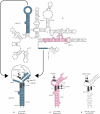
Figure 1.
Figure 1.
Secondary structure of the mRNA leader region of the rpmI-rplT operon. (A) Model of the secondary structure of the rpmI translational operator (13). infC, iris (an acronym for infC-rpmI intergenic sequence) and rpmI sequences are italicized, uppercased and lowercased, respectively. Residues are numbered according to infC, iris and rpmI numbering. Every 10th residue is marked with a tick mark and every 50th nucleotide is numbered. Some of the relevant features of the operator (stems S1, S2 and t1 containing the t1 transcriptional terminator) are also indicated. The nucleotides forming the rpmI Shine–Dalgarno sequence are indicated by asterisks. (B) Schematic view of the pseudoknotted site 1. infC and iris sequences and the nucleotides forming the rpmI Shine–Dalgarno sequence are indicated as in (A). The long-range base-pairing interaction which holds the pseudoknot is schematized by two converging arrows starting from the separated interacting partners in (A). (C) Schematic view of site 2. infC and iris sequences are indicated as in (A). (D) Schematic view of the H40-H41 junction of E. coli 23S rRNA. The names of the mutations used in the fluorescent measurement experiments are indicated by arrows pointing to the substituting nucleotides in the schematic representations of the three L20–RNA-binding sites.
Figure 2.
Figure 2.
Determination of the affinity of L20C-ter for its mRNA- and rRNA-binding sites. The fluorescence anisotropy titration curves for wild-type and mutant operator RNAs are shown in (A). The equivalent titration curves for the 23S rRNA are shown in (B) where the left and right ordinates are for anisotropy measurements performed with 23S rRNA and wild-type operator RNA, respectively.
Figure 3.
Figure 3.
Oligomerization state of L20C-ter: gel filtration analysis. The elution profile of the Superdex 75 HR column is shown with a continuous line for the exclusion volume (peak 1) and for the 3 standards (peaks 2, 3 and 4 corresponding to 44, 17 and 1.35 kDa, respectively). The elution profile of L20C-ter is shown with a dotted line. The calibration curve is shown in the insert where Log MW is the logarithm of the molecular weight and Kav = (Ve − Vd)/(Vt − Vd) where Ve is the elution volume, Vd the dead volume (8 ml), Vt the total volume (24 ml). The dotted line in the insert shows the Kav of L20C-ter from which its molecular weight was calculated.
Figure 4.
Figure 4.
Oligomerization state of L20C-ter: FCS measurements. L20C-ter-Alexa488 and L20C-ter-Atto647 were at a nominal concentration of 150 nM each. Open circles correspond to the autocorrelation profiles obtained from the data collected on channel 2 (corresponding to L20C-ter-Alexa488 emission) and dots correspond to the autocorrelation profiles calculated from the data collected in channel 1 (corresponding to L20C-ter-Atto647 emission). The crosses correspond to the cross-correlation profile between the fluctuations in channel 1 and channel 2. Buffer conditions are as described in the Materials and Methods section. Lines through the points correspond to the fits of the data as described in the text and for which the results are given in Table 1.
Figure 5.
Figure 5.
Affinity separation experiment. (A), (B), and (C) show RNA gels. In (A), wild-type operator RNA was adsorbed to the Co++-sepharose beads in the presence of His-L20C-ter (lanes1, 2, 3) or in its absence (lanes 4, 5, 6). FT, W, E stand for flow-through, wash and elution, respectively. In (B), His-L20C-ter was adsorbed to the Co++-sepharose beads in the presence of either wild-type (lanes 1, 2, 3) or mutant operator RNA (lanes 4, 5, 6). In (C), CBP-L20C-ter was adsorbed to the calmodulin-sepharose beads in the presence of either wild-type (lanes 1, 2, 3) or mutant operator RNA (lanes 4, 5, 6). (D) shows a protein gel where His-L20C-ter and CBP-L20C-ter were adsorbed to the Co++-sepharose beads in the absence (lanes 1, 2, 3) or presence (lanes 4, 5, 6) of wild-type operator RNA.
Figure 6.
Figure 6.
FCS measurements. Combinations of L20C-ter-Alexa488, L20-ter-Atto647 and operator RNA at a nominal concentration of 150 nM each were used. (A) and (B) Normalized autocorrelation profiles for channel 2 (L20C-ter-Alexa488) and channel 1 (L20C-ter-Atto647). Dots correspond to the protein alone in (A) L20C-ter-Alexa488 and (B) L20C-ter-Atto647. Triangles correspond to data obtained after the addition of the RNA. Crosses correspond to the autocorrelation profile obtained after addition of 150 nM of (A) L20C-ter-Atto647 to the L20C-ter-Alexa488/RNA complex and (B) L20C-ter-Alexa488 to the L20C-ter-Atto647/RNA complex. (C) and (D) auto correlation profiles from channel 1 (dots), channel 2 (open circles) and cross correlation profiles (crosses) obtained for the solutions in which (C) 150 nM of L20C-ter-Atto647 was added to the L20C-ter-Alexa488/RNA complex at a concentration of 150 nM and (D) 300 nM of L20C-ter-Atto647 was added to the L20C-ter-Alexa488/RNA complex at a concentration of 300 nM. Buffer conditions are as described in the Materials and Methods section. Lines through the points correspond to the fits of the data as described in the text and for which the results are given in Table 1.
Figure 7.
Figure 7.
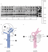
Localization of the nucleotide residues displaying increased reactivities towards chemical probes upon mutations in either site 1 or site 2. (A) Gel view showing the result of cDNA extension analysis spanning the infC318-infC354 region of the operator RNA. Extension analysis was performed using DEL2 oligodeoxynucleotide on modified wild-type (WT) or mutant (1: 335infC, 2: 78SIR, 3: iris1AA and 4: iris53-55) operator RNA. The chemicals (DMS, KET and CMCT) used for modification are shown beneath the corresponding lanes. Lane −, unmodified operator; lane +, modified operator. The primary structure of the infC318-infC354 region of the operator is shown on the right of the gel. U, G, C, A are sequencing lanes. Bands corresponding to increased reactivities induced by long-range destabilization and position of the corresponding modified nucleotide in the primary structure shown on the right of the gel view are indicated by filled triangles. (B) Schematic view of the pseudoknotted site 1. infC and iris sequences and the nucleotides forming the rpmI Shine–Dalgarno sequence are indicated as in the legend to Figure 1. The positions and substituting nucleotides of mutations 1 and 2 are indicated by arrows. Residues displaying increased reactivities due to local destabilization are within squares whereas the residue showing increased reactivity due to long-range destabilization is within circle and marked with a large star. The mutations inducing increased reactivities are indicated by their numbers in parenthesis. (C) Schematic view of site 2. infC and iris sequences are indicated as in the legend to Figure 1. The positions and substituting nucleotides of mutations 3 and 4 are indicated by arrows. Residues displaying and mutations inducing increased reactivities are indicated as in B.
Figure 8.
Figure 8.
Models of L20 binding to its operator. (A) L20 binding to H2/L1 hybrid site produced by interaction between a region of site 2 (H2) and a region of site 1 (L1). In this model, the remaining regions of both sites are unable to form a L20-binding site. (B) L20 binding to one preformed site excludes binding to the second site either by structural change of the latter or by steric hindrance. (C) L20 binding to site 2 induces a structural change of site 1 rendering the latter competent for L20 binding and/or local increase of L20 concentration which increases the L20-binding efficiency of site 1.

Similar articles

Cited by

  • Probing ribosomal protein-RNA interactions with an external force.
    Mangeol P, Bizebard T, Chiaruttini C, Dreyfus M, Springer M, Bockelmann U. Mangeol P, et al. Proc Natl Acad Sci U S A. 2011 Nov 8;108(45):18272-6. doi: 10.1073/pnas.1107121108. Epub 2011 Oct 24. Proc Natl Acad Sci U S A. 2011. PMID: 22025688 Free PMC article.
  • Regulation of translation initiation by RNA binding proteins.
    Babitzke P, Baker CS, Romeo T. Babitzke P, et al. Annu Rev Microbiol. 2009;63:27-44. doi: 10.1146/annurev.micro.091208.073514. Annu Rev Microbiol. 2009. PMID: 19385727 Free PMC article. Review.
  • Discovery of functional elements in 12 Drosophila genomes using evolutionary signatures.
    Stark A, Lin MF, Kheradpour P, Pedersen JS, Parts L, Carlson JW, Crosby MA, Rasmussen MD, Roy S, Deoras AN, Ruby JG, Brennecke J; Harvard FlyBase curators; Berkeley Drosophila Genome Project; Hodges E, Hinrichs AS, Caspi A, Paten B, Park SW, Han MV, Maeder ML, Polansky BJ, Robson BE, Aerts S, van Helden J, Hassan B, Gilbert DG, Eastman DA, Rice M, Weir M, Hahn MW, Park Y, Dewey CN, Pachter L, Kent WJ, Haussler D, Lai EC, Bartel DP, Hannon GJ, Kaufman TC, Eisen MB, Clark AG, Smith D, Celniker SE, Gelbart WM, Kellis M. Stark A, et al. Nature. 2007 Nov 8;450(7167):219-32. doi: 10.1038/nature06340. Nature. 2007. PMID: 17994088 Free PMC article.

References

    1. Romby P., Springer M. Translational control in prokaryotes. In: Matthews M.B., Sonenberg N., Hershey J.W.B., editors. Translational Control of Gene Expression. Cols Spring Harbor, Newyork: Cold Spring Harbor Laboratory Press; 2006.
    1. Hattman S. Unusual transcriptional and translational regulation of the bacteriophage Mu mom operon. Pharmacol. Ther. 1999;84:367–388. - PubMed
    1. Oppenheim A.B., Kornitzer D., Altuvia S., Court D.L. Posttranscriptional control of the lysogenic pathway in bacteriophage lambda. Prog. Nucleic Acid Res., Mol. Biol. 1993;46:37–49. - PubMed
    1. Zengel J.M., Lindahl L. Diverse mechanisms for regulating ribosomal protein synthesis in Escherichia coli. Prog. Nucleic Acid Res. Mol. Biol. 1994;47:331–370. - PubMed
    1. Nomura M., Gourse R., Baughman G. Regulation of the synthesis of ribosomes and ribosomal components. Annu. Rev. Biochem. 1984;53:75–117. - PubMed

Publication types

MeSH terms