A periodic pattern of mRNA secondary structure created by the genetic code
- PMID: 16682450
- PMCID: PMC1458515
- DOI: 10.1093/nar/gkl287
A periodic pattern of mRNA secondary structure created by the genetic code
Abstract
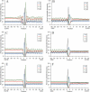
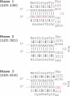
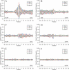
Single-stranded mRNA molecules form secondary structures through complementary self-interactions. Several hypotheses have been proposed on the relationship between the nucleotide sequence, encoded amino acid sequence and mRNA secondary structure. We performed the first transcriptome-wide in silico analysis of the human and mouse mRNA foldings and found a pronounced periodic pattern of nucleotide involvement in mRNA secondary structure. We show that this pattern is created by the structure of the genetic code, and the dinucleotide relative abundances are important for the maintenance of mRNA secondary structure. Although synonymous codon usage contributes to this pattern, it is intrinsic to the structure of the genetic code and manifests itself even in the absence of synonymous codon usage bias at the 4-fold degenerate sites. While all codon sites are important for the maintenance of mRNA secondary structure, degeneracy of the code allows regulation of stability and periodicity of mRNA secondary structure. We demonstrate that the third degenerate codon sites contribute most strongly to mRNA stability. These results convincingly support the hypothesis that redundancies in the genetic code allow transcripts to satisfy requirements for both protein structure and RNA structure. Our data show that selection may be operating on synonymous codons to maintain a more stable and ordered mRNA secondary structure, which is likely to be important for transcript stability and translation. We also demonstrate that functional domains of the mRNA [5'-untranslated region (5'-UTR), CDS and 3'-UTR] preferentially fold onto themselves, while the start codon and stop codon regions are characterized by relaxed secondary structures, which may facilitate initiation and termination of translation.
Figures


Similar articles
-
Comparative analysis of orthologous eukaryotic mRNAs: potential hidden functional signals.Nucleic Acids Res. 2004 Mar 18;32(5):1774-82. doi: 10.1093/nar/gkh313. Print 2004. Nucleic Acids Res. 2004. PMID: 15031317 Free PMC article.
-
RNA secondary structure profiling in zebrafish reveals unique regulatory features.BMC Genomics. 2018 Feb 15;19(1):147. doi: 10.1186/s12864-018-4497-0. BMC Genomics. 2018. PMID: 29448945 Free PMC article.
-
Multiple RNA structures affect translation initiation and UGA redefinition efficiency during synthesis of selenoprotein P.Nucleic Acids Res. 2017 Dec 15;45(22):13004-13015. doi: 10.1093/nar/gkx982. Nucleic Acids Res. 2017. PMID: 29069514 Free PMC article.
-
A code within the genetic code: codon usage regulates co-translational protein folding.Cell Commun Signal. 2020 Sep 9;18(1):145. doi: 10.1186/s12964-020-00642-6. Cell Commun Signal. 2020. PMID: 32907610 Free PMC article. Review.
-
A Code Within a Code: How Codons Fine-Tune Protein Folding in the Cell.Biochemistry (Mosc). 2021 Aug;86(8):976-991. doi: 10.1134/S0006297921080083. Biochemistry (Mosc). 2021. PMID: 34488574 Free PMC article. Review.
Cited by
-
Recent Findings on Therapeutic Cancer Vaccines: An Updated Review.Biomolecules. 2024 Apr 21;14(4):503. doi: 10.3390/biom14040503. Biomolecules. 2024. PMID: 38672519 Free PMC article. Review.
-
Probing RNA structural landscapes across Candida yeast genomes.Front Microbiol. 2024 Feb 26;15:1362067. doi: 10.3389/fmicb.2024.1362067. eCollection 2024. Front Microbiol. 2024. PMID: 38468856 Free PMC article.
-
Fitness benefits of a synonymous substitution in an ancient EF-Tu gene depend on the genetic background.J Bacteriol. 2024 Feb 22;206(2):e0032923. doi: 10.1128/jb.00329-23. Epub 2024 Jan 30. J Bacteriol. 2024. PMID: 38289064 Free PMC article.
-
Tailor made: the art of therapeutic mRNA design.Nat Rev Drug Discov. 2024 Jan;23(1):67-83. doi: 10.1038/s41573-023-00827-x. Epub 2023 Nov 29. Nat Rev Drug Discov. 2024. PMID: 38030688 Review.
-
Natural variation in codon bias and mRNA folding strength interact synergistically to modify protein expression in Saccharomyces cerevisiae.Genetics. 2023 Aug 9;224(4):iyad113. doi: 10.1093/genetics/iyad113. Genetics. 2023. PMID: 37310925 Free PMC article.
References
-
- White H.B., III, Laux B.E., Dennis D. Messenger RNA structure: compatibility of hairpin loops with protein sequence. Science. 1972;175:1264–1266. - PubMed
-
- Ball L.A. Secondary structure and coding potential of the coat protein gene of bacteriophage MS2. Nature New Biol. 1973;242:44–45. - PubMed
-
- Fitch W.M. The large extent of putative secondary nucleic acid structure in random nucleotide sequences or amino acid derived messenger-RNA. J. Mol. Evol. 1974;3:279–291. - PubMed
-
- Lagunez-Otero J., Trifonov E.N. mRNA periodical infrastructure complementary to the proof-reading site in the ribosome. J. Biomol. Struct. Dyn. 1992;10:455–464. - PubMed
-
- Ikemura T. Codon usage and tRNA content in unicellular and multicellular organisms. Mol. Biol. Evol. 1985;2:13–34. - PubMed
Publication types
MeSH terms
Substances
Grants and funding
LinkOut - more resources
Full Text Sources
Other Literature Sources


