Distribution of HPV infection and tumour markers in cervical intraepithelial neoplasia from cone biopsies of Mozambican women
- PMID: 15623485
- PMCID: PMC1770547
- DOI: 10.1136/jcp.2004.020552
Distribution of HPV infection and tumour markers in cervical intraepithelial neoplasia from cone biopsies of Mozambican women
Abstract
Aims: To evaluate human papillomavirus (HPV) infection in whole cervical cone specimens with cervical intraepithelial neoplasia (CIN). In addition, to evaluate the relation between the presence of CIN lesions and HPV infection and the expression of Ki-67, p53, cytokeratins, Gp230 glycoprotein, and simple mucin-type carbohydrates.
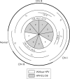
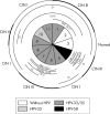
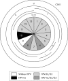
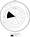
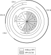
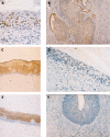
Methods: Cervical cone specimens from five patients with CIN were studied. For each specimen, serial sections encompassing the whole cone were collected (52 samples). HPV infection and HPV types were detected by the polymerase chain reaction and enzyme immunoassay. The expression of Ki-67, p53, cytokeratins, Gp230, and simple mucin-type carbohydrates was examined immunohistochemically.
Results: All cases showed high risk HPV types, namely types 16, 33, 35, and 58. Four of the five patients were infected by multiple viral types. HPV-58 was always seen in CIN III, whereas HPV-35 was more frequent in CIN I. The expression of Ki-67 and p53 was higher in CIN III lesions. The expression of cytokeratins 8 and 17 showed complete or almost complete overlap with CIN III. Altered expression of Gp230, Tn, and sialyl-T was often seen in all grades of CIN.
Conclusions: When whole cervical cone specimens are evaluated the rate of multiple HPV infection is very high. The expression of cytokeratins 8 and 17 is a useful marker of CIN III.
Figures
Similar articles
-
Aberrant expression of VEGF-C is related to grade of cervical intraepithelial neoplasia (CIN) and high risk HPV, but does not predict virus clearance after treatment of CIN or prognosis of cervical cancer.J Clin Pathol. 2006 Jan;59(1):40-7. doi: 10.1136/jcp.2005.026922. J Clin Pathol. 2006. PMID: 16394279 Free PMC article.
-
Characterization of human papillomavirus infection, P53 and Ki-67 expression in cervix cancer of Mozambican women.Pathol Res Pract. 2003;199(5):303-11. doi: 10.1078/0344-0338-00422. Pathol Res Pract. 2003. PMID: 12908520
-
Low- and high-risk CIN 1 and 2 lesions: prospective predictive value of grade, HPV, and Ki-67 immuno-quantitative variables.J Pathol. 2003 Apr;199(4):462-70. doi: 10.1002/path.1316. J Pathol. 2003. PMID: 12635137
-
Epidemiology of cervical intraepithelial neoplasia: the role of human papillomavirus.Baillieres Clin Obstet Gynaecol. 1995 Mar;9(1):1-37. doi: 10.1016/s0950-3552(05)80357-8. Baillieres Clin Obstet Gynaecol. 1995. PMID: 7600720 Review.
-
Aetiology, pathogenesis, and pathology of cervical neoplasia.J Clin Pathol. 1998 Feb;51(2):96-103. doi: 10.1136/jcp.51.2.96. J Clin Pathol. 1998. PMID: 9602680 Free PMC article. Review.
Cited by
-
Correlation of P16 (Ink4a) and CK17 to HPV (16E6+18E6) in Premalignant and Malignant Lesions of Uterine Cervix: A Clinicopathologic Study.Iran J Pathol. 2016 Fall;11(4):377-390. Iran J Pathol. 2016. PMID: 28855930 Free PMC article.
-
Human papillomavirus prevalence and genotype distribution among young women and men in Maputo city, Mozambique.BMJ Open. 2017 Jul 17;7(7):e015653. doi: 10.1136/bmjopen-2016-015653. BMJ Open. 2017. PMID: 28716790 Free PMC article.
-
Human papillomavirus (HPV) infection: a Mozambique overview.Virusdisease. 2016 Jun;27(2):116-22. doi: 10.1007/s13337-016-0319-7. Epub 2016 May 3. Virusdisease. 2016. PMID: 27366761 Free PMC article. Review.
-
Prevalence and risk factors for cancer of the uterine cervix among women living in Kinshasa, the Democratic Republic of the Congo: a cross-sectional study.Infect Agent Cancer. 2015 Jul 15;10:20. doi: 10.1186/s13027-015-0015-z. eCollection 2015. Infect Agent Cancer. 2015. PMID: 26180542 Free PMC article.
-
Trends in cancer incidence in Maputo, Mozambique, 1991-2008.PLoS One. 2015 Jun 25;10(6):e0130469. doi: 10.1371/journal.pone.0130469. eCollection 2015. PLoS One. 2015. PMID: 26110774 Free PMC article.
References
-
- Carrilho C , Gouveia P, Cantel M, et al. Characterization of human papillomavirus infection, P53 and Ki-67 expression in cervix cancer of Mozambican women. Pathol Res Pract 2003;199:303–11. - PubMed
-
- Castellsague X , Menendez C, Loscertales MP, et al. Human papillomavirus genotypes in rural Mozambique. Lancet 2001;358:1429–30. - PubMed
Publication types
MeSH terms
Substances
LinkOut - more resources
Full Text Sources
Medical
Research Materials
Miscellaneous

